Introduction
Lower risk and occurrence of chronic diseases have been correlated with whole grains of cereals, legumes and pseudocereals regular intake (Ayyash et al., 2019; Malaguti et al., 2014). Therefore, there is a need to understand the attributes of legumes and to obtain specific components with functional properties without extensive processing (Barbana & Boye, 2010). Protein derived hydrolysates may represent one such source of health-enhancing components.
Recently, bioactivity of protein hydrolysates has been reported, including antihypertensive, antioxidant, immunomodulatory, opioid and hypercholesterolemic activities (Aluko, 2019; Ayyash, Johnson, Liu, Al-Mheiri, & Abushelaibi, 2018; Lammi, Aiello, Boschin, & Arnoldi, 2019).
Several research studies have shown that enzymatic hydrolysates contain bioactive peptides, which are small amino acids sequences released from native proteins (NP) by hydrolysis. Protein hydrolysates from soy, lentil, bean, chickpea, and pea have received much attention (Hernández-Álvarez et al., 2013; Roy, Boye, & Simpson, 2010). In contrast, there are very scarce studies about protein hydrolysates from lupin (Lupinus gredensis), which has a protein content comparable to that of soybean (Ayyash et al., 2019; Vioque, Alaiz, & Girón-Calle, 2012).
Karkouch et al. (2017) identified peptides with antioxidant, antityrosinase and antibiofilm activities released from faba bean seed protein by trypsin, which could be potentially used in cosmetic and pharmaceutical industries. Jakubczyk et al. (2019) inform that the conditions of the faba bean seed fermentation process have an influence on the molecular mass of proteins and activities of peptides released from the protein during in vitro digestion.
Protein hydrolysates are mostly produced by in vitro enzymatic hydrolysis (EH) using food-grade enzyme preparations or by fermentation using microorganisms as a source of proteolytic enzymes (Chibuike & Aluko, 2012). The latter process is a simpler and more economical means of obtaining protein hydrolysates compared to EH (Ferri, Serrazanetti, Tassoni, Baldissarri, & Gianotti, 2016; Marques et al., 2019). Both methods produce protein hydrolysates that differ in structural and functional characteristics (Chibuike & Aluko, 2012), so that it is of the utmost importance to gain understanding about the factors affecting the biological properties of these components.
It is difficult to translate the in vitro health-promoting effects of the protein hydrolysates on human as bioactive peptides may be degraded during digestion. It is, therefore, important to have information regarding the resistance of the protein hydrolysates to gastrointestinal enzymes (Carbonaro, Maselli, & Nucara, 2015). Based on the above the objectives of this study were to: 1) produce bioactive hydrolysates from lupin and faba bean proteins by EH and solid-state fermentation (SF), 2) compare the angiotensin converting enzyme (ACE)-inhibitory and antioxidant activities of the hydrolysates, and 3) evaluate the effect of in vitro gastrointestinal digestion on the antioxidant and antihypertensive activities of the hydrolysates.
Materials and methods
Seeds
Faba bean (F; Vicia faba) and lupin (L; Lupinus gredensis) seeds were purchased from Empacadora de Semillas Zaragoza, S.A. de C.V. (Mexico City, Mexico). Flours were obtained by milling seeds in a cutter mill (modelo de plástico, Pulvex®, Mexico), and the fractions that were retained between 0.14 and 1.19 mm sieves were collected. Flours were vacuum packed in polyethylene bags, and stored under dark conditions at 4 °C. Flours were analysed for protein, moisture, ash, crude fiber, and fat (Association of Official Analytical Chemists [AOAC], 2005). The remaining percentage was considered to represent carbohydrates.
Materials
Aspergillus niger GH1 strain was obtained from Food Research Department, University of Coahuila (GenBank: HQ450381.1). Flavourzyme® 1000 L (EC 3.4.11.1, a mixture of endoproteinase and exopeptidase of Aspergillus oryzae; 1 000 LAPU·g-1) was purchased from Novo Nordisk (Denmark). Potato dextrose agar (PDA) was purchased from DIFCO™ (USA). Tween-80, 2,4,6-trinitrobenzenesulfonic acid (TNBS), L-leucine, tannic acid, 2,2-diphenyl-1-picrylhydrazyl (DPPH), hippuryl-L-histidyl-L-leucine (HHL), hippuric acid (HA), Hammarsten grade casein, pepsin (P7000), pancreatin (P1750), angiotensin converting enzyme (ACE-I, A6778), and Whatman No. 41 filter paper were purchased from Sigma-Aldrich® (USA). Electrophoresis reagents were obtained from Bio-Rad® (USA). All other chemicals used in the experiments were of analytical grade, and all the water used was double distilled and deionized (DDW).
Enzymatic hydrolysis (EH)
Protein extraction from the flours was carried out according to Alsohaimy, Sitohy, and El-Masry (2007) with slight modifications. Flour samples (50 g) were suspended in 1 L of DDW. pH was adjusted to 12 with 6 N and 0.1 N NaOH, and the suspensions were stirred at 100 rpm using an orbital shaker (E-2500, Thermo Fisher®, USA), for 120 min at 25 °C. Afterwards, 1 mL samples were taken and centrifuged (X-12R, Allegra®, USA) at 6 000 g for 30 min at 4 °C. Soluble protein in the supernatants was determined by the modified Lowry method (Peterson, 1977).
Supernatants were poured into Erlenmeyer flasks, pH was adjusted to 7 using 6 N and 0.1 N HCl, and Flavourzyme® 1000 L in an enzyme to substrate (E/S) ratio of 125 LAPU·gprotein -1 was added. The Erlenmeyer flasks were continuously stirred (150 rpm, 8 h, 50 °C). Aliquots (250 µL) were withdrawn every 60 min, added with 0.01 g·mL-1 sodium dodecyl sulfate, and heated at 85 °C for 15 min to inactivate the enzymes mix. Hydrolysates were centrifuged (10 000 g, 30 min, 4 °C) and filtered to remove insoluble residues. The filtrates were used for the determination of degree of hydrolysis (DH percentage) and molecular weight (MW) distribution, freeze-dried (Lyph Lock 6, Labconco, USA) and stored at -20 °C until required for antioxidant and ACE-inhibitory analysis. F and L enzymatic hydrolysates were coded as EHF and EHL, respectively.
Solid-state fermentation (SF) hydrolysis
The fungal strain was cultured for 5 days at 30 °C in Erlenmeyer flask (250 mL) with 50 mL of PDA. Spores were harvested using 10 mL of Tween 80 (0.1 % v/v). SF was carried out by placing 10 g of each flour in 250 mL Erlenmeyer flasks. The moisture content was adjusted at 50 % (w/w) with a salts solution (pH 6) containing (% w/v): dipotassium hydrogen phosphate (0.1), magnesium sulphate (0.005) and potassium chloride (0.005). Flasks were autoclaved at 121 °C for 20 min, cooled to 30 °C, inoculated with 2 × 10⁷ spores·g-1 of dried flours, and incubated at 30 °C. Samples (duplicates) were withdrawn at 24 h intervals during 6 days, added with DDW (1:3), vortexed for 1 min, and heated at 85 °C for 15 min to inactivate the proteases. The insoluble material was removed by centrifugation at 10 000 g, for 10 min at 4 °C and filtered. Filtrates were used for the determination of DH percentage and MW distribution analysis, then freeze-dried and stored at -20 °C, until required for analysis. In the case of protease activity determination samples were withdrawn before heating at 85 °C. F and L hydrolysates were coded as SFF and SFL, respectively.
Degree of hydrolysis
DH percentage was calculated by determining free amino groups through reaction with TNBS using a leucine standard (Addler-Nissen, 1979). The total number of amino groups was determined in a 100 % hydrolyzed sample treated with 6 N HCl at 110 °C for 24 h in a vacuum oven (Memmert, USA).
Fungal protease activity
The fungal protease activity in the SF hydrolysates was determined according to the method reported by Hernández-Martínez et al. (2011) with slight modifications. Briefly, the reaction mixture consisted of 570 µL of a solution containing Hammerstein grade casein at 0.01 g·mL-1 in phosphate buffer (50 mM, pH 7) pre-incubated at 50 °C for 5 min and 30 µL of SF hydrolysate. The enzymatic reaction was performed during 15 min and stopped by adding 0.9 mL of trichloroacetic acid (TCA; 0.050 g·mL-1). The mixture was centrifuged at 10 000 g for 15 min at 4 °C, and filtered. Soluble peptides in TCA were estimated by the modified Lowry method. One enzyme unit (U) was defined as the amount of enzyme required to produce an increment of 0.01 in absorbance at 750 nm under the assay conditions (Soares-de Castro & Sato, 2014). Enzyme activity was expressed in U·per gram of dry matter (U·g-1) (Castañeda-Casasola et al., 2018).
Electrophoresis
MW distribution of the NP and their hydrolysates was determined by SDS-polyacrylamide gel electrophoresis (SDS-PAGE) as described by Laemmli (1970) using a Mini-Protean 2 gel electrophoresis unit (Bio-Rad®, USA). Samples were dissolved in a 1:2 (v/v) ratio in a buffer solution: SDS (20 % w/v), glycerol (2.5 % v/v), β-mercaptoethanol (1.0 % v/v), and bromophenol blue (0.02 % w/v). Resolving gel (pH 8.8) consisted of SDS (0.21 % w/v), and acrylamide (20 % w/v). Stacking gel (pH 6.8) was made up of SDS (0.21 % w/v) and acrylamide (5 % w/v). The total gel thickness was 0.75 mm, with 10 cm of resolving gel and 2 cm of stacking gel. Protein bands were stained by gel immersion in Coomassie brilliant blue R-250 (Cat. 161-0400, Bio-Rad®) in a solution of 400 mL of methanol, 70 mL of acetic acid and 530 mL of DDW (Hernández-Álvarez et al., 2013). Broad-range protein MW standards in the range from 7.1 to 103 kDa (Cat. 161-0303, Bio-Rad®) were used.
Antioxidant and ACE-inhibitory activities
The antioxidant and ACE-inhibitory activities of the proteins and their hydrolysates were determined at selected times. DPPH free radical scavenging activity was determined as described by Cheison, Wang, and Xu (2007) with slight modifications. Five serial dilutions (0 - 6 mg·mL-1 of protein) were dissolved in 0.05 M sodium carbonate/bicarbonate buffer (pH 9.6). Aliquots of 500 µL were mixed with 1.5 mL of DPPH (0.08 mM) in methanol (80 % v/v), allowed to stand (in dark) at room temperature for 60 min, and the reduction of the DPPH radical was measured at 517 nm (UV-160, Shimadzu®, Japan). The buffer was used as blank.
Antioxidant activity was expressed as the 50 % inhibition percentage (IC50) of the free radical DPPH, and calculated by regression analysis of DPPH inhibition (%) versus hydrolysate protein concentration (mg·mL-1).
The ACE-inhibitory activity assay was performed using the method reported by Cushman and Cheung (1971), with slight modifications. The reaction mixture contained 5 mM of hippuryl-L-histidyl-L-leucine as substrate, 0.3 M NaCl and 2 mU of enzyme in 50 mM sodium borate buffer (pH 8.3). A sample (100 μL) was added to reaction mixture (200 μL) and incubated at 37 °C for 45 min. The reaction was stopped by the addition of 1.0 N HCl (300 μL). Then 1 mL of ethyl acetate was added to the mixture, vortexed (30 s) and centrifuged at 6 000 g (4 °C, 10 min). The upper layer (750 µL) was transferred into a glass tube and evaporated at room temperature for 2 h in vacuum. The hippuric acid was re-dissolved in 1.6 mL of DDW, and absorbance was measured at 228 nm. The IC50 value was determined by regression analysis of ACE inhibition (%) versus hydrolysate protein concentration (0 to 15 mg·mL-1) of serial dilutions.
In vitro gastrointestinal digestion
Simulated gastrointestinal digestion was carried out by sequential pepsin and pancreatin action on the hydrolysates according to the method reported by Ketnawa, Martínez-Alvarez, Benjakul, and Rawdkuen (2016) with slight modifications. Freeze dried hydrolysates were resolubilized (3 % w/v) in DDW, adjusted to pH 2.0 with 1 N HCl, and added with pepsin (4 % w/w of protein). The mixture was incubated at 37 °C for 2 h. The pH was then adjusted to 7.5 with 1 N NaOH, and pancreatin was added (10 % w/w of protein). The resulting mixture was incubated at 37 °C for 2 h, and afterwards digestion was stopped by keeping the test tubes in boiling water for 10 min. The mixtures were cooled at room temperature and centrifuged at 10 000 g for 15 min. The supernatants were lyophilized, kept in plastic tubes and stored at -20 °C before antioxidant and ACE-inhibitory activities determination.
Data analysis
The data are presented as mean ± standard deviation. Analyzes were carried out in triplicate from three independent experiments. One-way analyses of variance were performed to evaluate the chemical composition of flours and the characteristics of hydrolysates, and Tukey (P ( 0.05) means comparison tests were performed. The SPSS statistical program (SPSS Inc., USA) was used for these tests.
Results and discussion
Proximate chemical composition of seed flours
Moisture, protein, carbohydrates, and ash contents were significantly different for the L and F seed flours. F had higher protein content than L (Table 1). Olukomaiya et al. (2020) reported higher protein (35.77 %) and fat (6.05 %) contents for L. angustifolius L. flour. Relatively high protein contents in mature seeds are due to protein accumulation throughout their development, varying slightly depending on plant species, variety, maturity and growing conditions.
Table 1 Chemical proximate composition of lupin and faba bean seed flours (g·100 g-1 dry basis).
| Chemical composition | Seed | |
|---|---|---|
| Lupin | Faba bean | |
| Protein | 22.2 ± 2.0 az | 30.0 ± 1.0 b |
| Fat | 1.4 ± 0.6 a | 1.6 ± 0.1 a |
| Carbohydrates | 58.6 ± 4.6 b | 50.2 ± 2.1 a |
| Fiber | 15.0 ± 2.0 a | 14.9± 0.1 a |
| Ash | 2.8 ± 0.1 a | 3.3 ± 0.3 b |
zMeans with the same letter within each row are not statistically different (Tukey, P ≤ 0.05).
Protein extraction
Protein extraction is affected by pH, which influences the ratio of free to neutralized charges (Alsohaimy et al., 2007). The pH values ( 8 promote tannin-protein dissociation which hampers the enzymatic reaction (Guang & Phillips, 2009). In this work, it was found that the use of alkaline solutions at pH 12 produced solubilized protein yields of 87.7 ± 0.9 % for L and 94.9 ± 2.8 % for F.
Degree of hydrolysis and protease activity
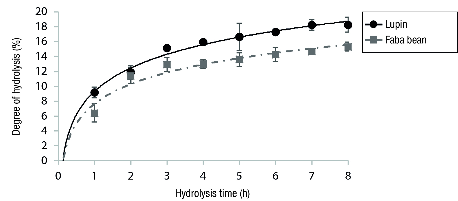
The extent of seed proteins hydrolysis was monitored using the DH percentage, which is defined as the percentage of the total number of peptide bonds in a protein which has been cleaved during hydrolysis (Addler-Nissen, 1979). The EH curves of F and L proteins were characterized by an initial pronounced increased in DH values (up to approximately 1 h) (Figure 1). Afterwards the hydrolysis rate decreased (around 2-6 h) and finally approached to a quasi-stationary state, where very slow hydrolysis occurred possible due to exhaustion of the substrate, enzyme inhibition by products or autolysis (Kristinsson & Rasco, 2000). This typical EH profile has been also reported for proteins from mung-bean and black bean var. Jamapa (Hernández-Álvarez et al., 2013; Hong, Wei, Liu, & Hui, 2005).
The DH values obtained after 6 h of EH were of 15.3 ± 0.2 % for F and of 18.3 ± 1.0 % for L. The DH percentage obtained for faba bean protein was higher than that reported for faba bean protein hydrolyzed with Flavourzyme during 1 h (Eckert et al., 2019). The different DH values shown by the seeds could be due to the differences in their storage protein profile, and the secondary and tertiary protein structures, which influenced their susceptibility to proteolysis (Hernández-Álvarez et al., 2013).

Figure 1 Degree of hydrolysis of lupin (L) and faba bean (F) proteins by Flavourzyme® 1000 L (125 LAPU·gprotein -1, 50 °C, pH 7).
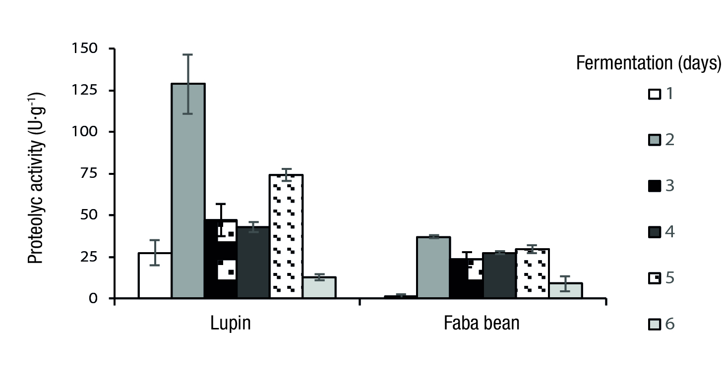
Protease production by SF was assessed with F and L flours as substrates for A. niger. The results showed an inconsistent pattern of protease production over the course of fermentation of both seeds. Figure 2 shows increasing levels of enzymatic activity up to 2 days reaching values of 37.03 ± 0.97 U·g-1 for F and 128.90 ± 17.75 U·g-1 for L. Afterwards protease production decreased during the following 3 to 4 days, but for the case of L a new increase of enzymatic activity was observed at 5 days. The DH values obtained after 6 days of fermentation were 6.9 ± 0.1 % for F and 8.6 ± 0.2 % for L.

Figure 2 Influence of the incubation period on protease production by Aspergillus niger GH1 under solid state fermentation using lupin and faba bean flours.
Novelli, Barros, and Fleuri (2016) reported that differences in protease production by SF might depend on either the type of substrate or the fungal strain. They also reported that similar protease production for more than one fermentation time could occur, and that the variations in proteolytic activity with fermentation time could reflect underlying adaptations in the molecular and physiological machinery of a particular strain for a given substrate. In the particular case of SF by A. niger, they found a maximum proteolytic activity for wheat bran after 4 days and for soybean bran after 5 days. Furthermore, the knowledge of the microbial biological mechanisms associated with this process helped to achieve optimal levels of enzyme production by SF. Belmessikh, Boukhalfa, Mechakra-Maza, Gheribi-Aoulmi, and Amrane (2013) found various fermentation times for A. oryzae according to the proteolytic activity measured and the substrate used.
On the other hand, Sumantha, Sandhya, Szakacs, Soccol, and Pandey (2005) reported that the drop in enzyme activity with increasing fermentation time could be due to cessation of production, or to inactivation of the proteases by autolysis. Proteases participate in many essential general processes of cells and their regulation. The simplest and most obvious role of microbial proteases is in nutrition where extracellular proteases degrade insoluble proteins and soluble large polypeptides into smaller peptides and amino acids, which are accessible to the cell (Ward, Rao, & Kulkarni, 2009), and stimulate enzyme production (Chutmanop, Chuichulcherm, Chisti, & Srinophakun, 2008).
Molecular weight distribution
The MW distribution of the NP and hydrolysates was examined by SDS-PAGE (Figure 3). Samples processed for 120 h in the case of SF and for 6 h for EH were analyzed, as longer times did not produce major differences in the polypeptide profiles.

Figure 3 SDS-PAGE of native protein (NP), fermented protein hydrolysates (SF) and enzymatic protein hydrolysates (EH) of: a) lupin (L) and b) faba bean (F). Lane St: MW standards (kDa).
SDS-PAGE of NP of L (NPL) and F (NPF) (Figures 3a and 3b) showed bands corresponding to α’/α (~77 kDa) and β (~48 kDa) subunits of 7S (β-conglycinin), convicilin (63 kDa), and acid and basic subunits of 11S globulin (~30-43 kDa and 20-25 kDa, respectively). It has been reported that the bands between 34-37 kDa and 18-25 kDa of faba bean protein correspond to the acidic and basic subunits of the 11S fraction, respectively (Eckert et al., 2019). Also low MW (< 20.7 kDa) bands can be observed that could be due to a mixture of albumin polypeptides, γ-vicilin or polypeptides from the post-translational cleavage of the storage proteins (Barbana & Boye, 2010).
SFL showed the disappearance of the bands corresponding to β-conglycinin, convicilin and acid subunits of 11S globulin by the proteolytic action of the proteases produced by A. niger, and released peptides exhibiting MW´s lower than 20.7 kDa (Figure 3a). Meanwhile EHL presented disappearance of the bands corresponding to α’/α subunits of 7S (β-conglycinin) and convicilin, and the production of peptides with predominantly MW´s at ≤ 7.1 kDa. SFF (Figure 3b) displayed decreases in the intensity of the major proteins bands and the appearance of bands corresponding to peptides with MW´s around of 40, 28.8, 20.7, and lower than 7.1 kDa. The basic subunits of 11S globulin were resistant to hydrolysis by A. niger proteases. On the other hand, EHF (Figure 3b) showed mainly peptides of MW’s < 7.1 kDa, so it may be inferred that EH was more efficient than SF.
Antioxidant and ACE-inhibitory activities
DPPH is a stable free radical and shows maximum absorbance at 517 nm in methanol. When DPPH encounters a proton-donating substance such as an antioxidant, the radical would be scavenged and the absorbance is reduced. Therefore, DPPH is widely used to evaluate the free radical scavenging activity of natural antioxidants (Wang, Le, Shi, & Zeng, 2014).
NPL and NPF did not show free radical scavenging ability, while all the hydrolysates showed antioxidant activity (Table 2). EHL showed a significantly higher antioxidant activity than the rest of the hydrolysates, which were non-significant different among themselves (Table 2). These results indicate that antioxidant activity was not directly related to the DH value or to the MW of the hydrolysates. Jakubczyk et al. (2019) indicated that not only short peptides consisting in lower than 20 amino acids present biological activity. Chen, Muramoto, Yamauchi, Fujimoto, and Nokihara (1998) concluded that the antioxidant properties of peptides are more related to their composition, structure, and hydrophobicity.
Table 2 Antioxidant and ACE-inhibition activities of the lupin (L) and faba bean (F) protein hydrolysates before and after in vitro gastrointestinal digestion.
| Hydrolyzate type | Undigested | Digested | Undigested | Digested | |
|---|---|---|---|---|---|
| DPPH IC50 (mg·mL-1) | ACE IC50 (mg·mL-1) | ||||
| EHL | 1.23 ± 0.02 az | 4.76 ± 0.10 b | 2.39 ± 0.10 a | 8.14 ± 0.53 a | |
| SFL | 1.97 ± 0.11 b | 33.86 ± 4.36 d | 14.08 ± 2.21 b | 12.25 ± 1.09 b | |
| EHF | 2.04 ± 0.10 b | 5.49 ± 0.06 b | nd | 12.58 ± 0.99 b | |
| SFF | 2.08 ± 0.03 b | 9.60 ± 0.04 c | nd | 7.48 ± 1.15 a | |
IC = 50 % inhibition; DPPH = 2,2-diphenyl-1-picrylhydrazyl; ACE = angiotensin converting enzyme; EH = enzymatic hydrolysis; SF = solid-state fermentation hydrolysis; nd = undetected. zMeans with the same letter within each column are not statistically different (Tukey, P ≤ 0.05).
The antioxidant properties of peptides have been attributed to the presence of certain amino acids (His, Tyr, Trp, Met, Lys, Cys) and to their correct positioning in peptide sequence (Sarmadi & Ismail, 2010). Hydrolysis may increase or decrease the hydrophobicity of the peptides depending on the nature of the precursor protein and MW of the generated peptides (Calderón-de la Barca, Ruiz-Salazar, & Jara-Marini, 2000). Erdmann, Cheung, and Schröder (2008) attributed the antioxidant activity of peptides to high concentrations of histidine and hydrophobic amino acids, with Pro-His-His sequences. DPPH IC50 values of our hydrolysates were significantly lower than those for cotton seed peptides reported by Sun et al. (2014) and wheat germ peptides obtained by Niu, Jiang, y Pan (2013).
Only the EHL (2.39 mg·mL-1) and SFL (14.08 mg·mL-1) hydrolysates undigested presented ACE-inhibitory activity (Table 2). EHF and SFF presented lower values of DH than EHL and SFL. It has been informed a positive correlation between the inhibitory capacity of the ACE and the DH (Fajardo-Espinoza, Romero-Rojas, & Hernández-Sánchez, 2020). The IC50 values of EHL and SFL were higher than those of hydrolysates of L. albus and L. angustifolius (0.226-0.268 mg·mL-1) obtained by Boschin, Scigliuolo, Resta, and Arnoldi (2014) with pepsin, probably due to in this last case the ACE-inhibitory peptides were separated by membrane filtration.
Liu, Chen, and Lin (2005) reported that tripeptides composed of amino acids with strong hydrophobicity at their C- and N-terminal had potent ACE-inhibitory activity. Arnoldi, Boschin, Zanoni, and Lammi (2015) observed IC50 values of protein hydrolysates of L. albus, L. angostifolius and L. luteus obtained with different enzymes between 0.136-1.053 mg·mL-1.
The comparison of ACE IC50 values obtained by different authors is a complex task, since the observed differences in the ACE-inhibitory activity may be related to various factors, such as the protein extraction procedure, peptide mixture composition, hydrolysis parameters, hydrolysis agent, analytical method for determining ACE-inhibitory activity, among others (Chin et al., 2019).
In vitro gastrointestinal digestion
The resistance of bioactive peptides against gastrointestinal proteases is a pre-requisite for their action in vivo and potential use as functional ingredients. Peptides resistant to gastrointestinal digestion can be absorbed in their intact form through the intestine to reach their target sites (Hannelore, 2004). Hence, the importance of evaluating the resistance of hydrolysates obtained against digestive enzymes.
After digestion all the hydrolysates showed DH percentage increases as follows: 12.7 ± 1.3 % for EHL, 3.2 ± 0.4 % for SFL, 5.6 ± 1.4 % for EHF and 7.5 ± 1.9 % for SFF. These results suggested differences in the susceptibility of the hydrolysates to digestive enzymes, and caused modifications in their antioxidant and ACE-inhibitory activities (Table 2). Significant antioxidant activity reductions were observed in the digested hydrolysates, which showed higher DPPH IC50 values. Girgih et al. (2015) reported that non-fractioned hydrolysates show a greater antioxidant activity due to a synergistic effect between peptides of different MWs.
ACE-inhibitory activity of the digested EHL was significantly lower than that before digestion, while that of SFL remained without significant changes.
Conclusions
Lupin hydrolysates exhibited antioxidant and ACE-inhibitory activities, while the native protein did not exhibit neither of these activities. The enzymatic hydrolysis of lupin protein by Flavourzyme yielded hydrolysates with higher biological activities than those produced by solid-state fermentation by A. niger. faba bean hydrolysates only exhibited antioxidant activity.
The in vitro evaluation of the lupin and faba bean protein hydrolysates against digestive enzymes indicated that the antioxidant activity diminished for both, while the ACE-inhibitory activity decreased for the lupin enzymatic hydrolysate, but remained without changes for fermented lupin hydrolysate. Therefore, it can be said that EH and SF improve the health-promoting properties of native bean and lupine proteins; however, more research is needed to determine the relationship between structure and activity of these protein hydrolysates.


 texto en


