Introduction
With a total area of 109,886 km2, Cuba is the largest and most biologically rich archipelago in the Caribbean basin. It harbors more than 7,000 vascular plant species, approximately 6000 of which have flowers and 50% are endemic (Berazaín et al., 2005). There are about 30 vegetation types, including semi desert, dry forest and tropical rainforest (Vales et al., 1998). However, over 500 years, the area of forest cover has been reduced from 94% to 15%, with a consequent loss of known species and fragmentation of habitat (Berazaín et al., 2005).
With 30 threatened species, 14 categorized as Critically Endangered and 16 as Endangered, sensu IUCN, Coccothrinax (c. 54 species) is the flagship palm genus for conservation in the Caribbean Island Biodiversity Hotspot (Jestrow et al., 2017). According to the Cuban Flora Red List, which follows IUCN criteria, a total of 33 taxa of Coccothrinax (32 of them endemics) are threatened (González-Torres et al., 2016): 8 Vulnerable (VU), 12 Endangered (EN), and 13 Critically Endangered (CR), with C. crinita included in the latter (Johnson, 1996).
Coccothrinax crinita (Griseb., & H.Wendl. ex C.H.Wright) Becc. is strictly endemic to Bahía Honda municipality and is considered one of the most endangered palms in the insular territories of America (Johnson, 1996). The species, known as “old man palm” or “palma petate”, has been used to provide vegetal fiber for rope, brooms, mattress stuffing, hats, purses and for fence poles (Martínez & Miranda, 2009). Its sustainable use can contribute to the conservation of its population and ecosystem.
It is well known that preservation of tropical endangered species is the main goal of numerous conservation projects, as well as an obligation for a number of countries bound by international agreements (González-Torres et al., 2016). Therefore, there is an urgent need for applied interdisciplinary studies on plants of special concern in order to develop effective methods for the maintenance and propagation of these species.
Mycorrhizal symbiosis is distributed widely in the natural world, forming mutualistic associations with most terrestrial plants and helping them to obtain and utilize nutrients (Koide & Mosse, 2004). This symbiosis also helps the plants to cope with adverse conditions, such as drought and salinity, as well as protecting plants from rhizophagous nematodes and soil pathogens (de la Pena et al., 2006; Roldán et al., 2008; Sheng et al., 2008). In tropical forests where competition for light, nutrients and space is high, association with arbuscular mycorrhizal fungi (AMF) increases the ability of the plant to quickly absorb nutrients and thus enhances growth and adaptability (Allen et al., 2003; Zubek et al., 2009).
Mycorrhizal associations have been reported in several palm species through measurement mycorrhizal colonization levels (Carrillo et al., 2002; Janos, 1977; St. John, 1988; Wang & Qiu, 2006), but few studies have addressed the effects of the mycorrhizal fungi on their host species. Palms that have been studied to date include Cocos nucifera L. (Ambili et al., 2012), Desmoncus orthacanthos Martius (Ramos-Zapata et al., 2006a, b), Elaeis guineensis Jacq. (Corley & Tinker, 2003; Nadarajah, 1980), Roystonea spp. O.F. Cook (Zona, 1996) and Coccothrinax readii H.J.Quero (Polanco et al., 2013). In the rhizosphere of Desmoncus orthacanthos, around 400 to 3,200 spores were found per 100 g of soil (Ramos-Zapata et al., 2006b); in a coconut plantation in Kerala, India, this value ranged from 428 to 598 spores (Rajeshkumar et al., 2015), while in Bactris gasipaes Kunth in the Amazon, 27 to 48 spores were found in 50 cm3 of soil (da Silva-Júnior & Cardoso, 2006). However, as highlighted by Fisher and Jayachandran (1999), mycorrhizal associations in tropical palms have received relatively little attention.
Two general AMF anatomical types have been described: Arum and Paris (Dickson et al., 2007; Gallaud, 1905; Smith & Smith, 1997). However, the factors that determine the formation of these types are not well understood and there is contradictory information that indicates host control, fungal control, or even control by both partners (Cavagnaro et al., 2001; Dickson, 2004; Dreyer et al., 2010; van Aarle et al., 2005). These AMF anatomy types have been described for some palm species as follows: a) Paris-type: E. guineensis (Nadarajah, 1980) and D. orthacanthos (Ramos-Zapata et al., 2006a, b), and b) Arum type: Acoelorraphe wrightii (Griseb. et Wendl.) Wendl. ex Becc., Coccothrinax argentata León, Phoenix dactylifera L., Pseudophoenix sargentii Wendl., Sabal palmetto (Walt.) Lodd. ex Schult. et Schult. F., Serenoa repens (Bartram) Small, and Thrinax morrisii Wendl. (Bouamri et al., 2006; Fisher & Jayachandran, 1999, 2005).
The first step in implementing strategies of conservation and restoration is to acquire the knowledge of mutualistic interactions that can increase success in plant establishment and survival. Quantifying the presence of fungi within the roots and spores in the rhizosphere contributes to this goal. An understanding of the AMF relationships of C. crinita can contribute to conservation of the species and facilitate its cultivation for economic purposes. The objective of the present work was, therefore, to determine the mycorrhizal status of C. crinite, as well as the density and richness of AMF spores present in its rhizosphere.
Materials and Methods
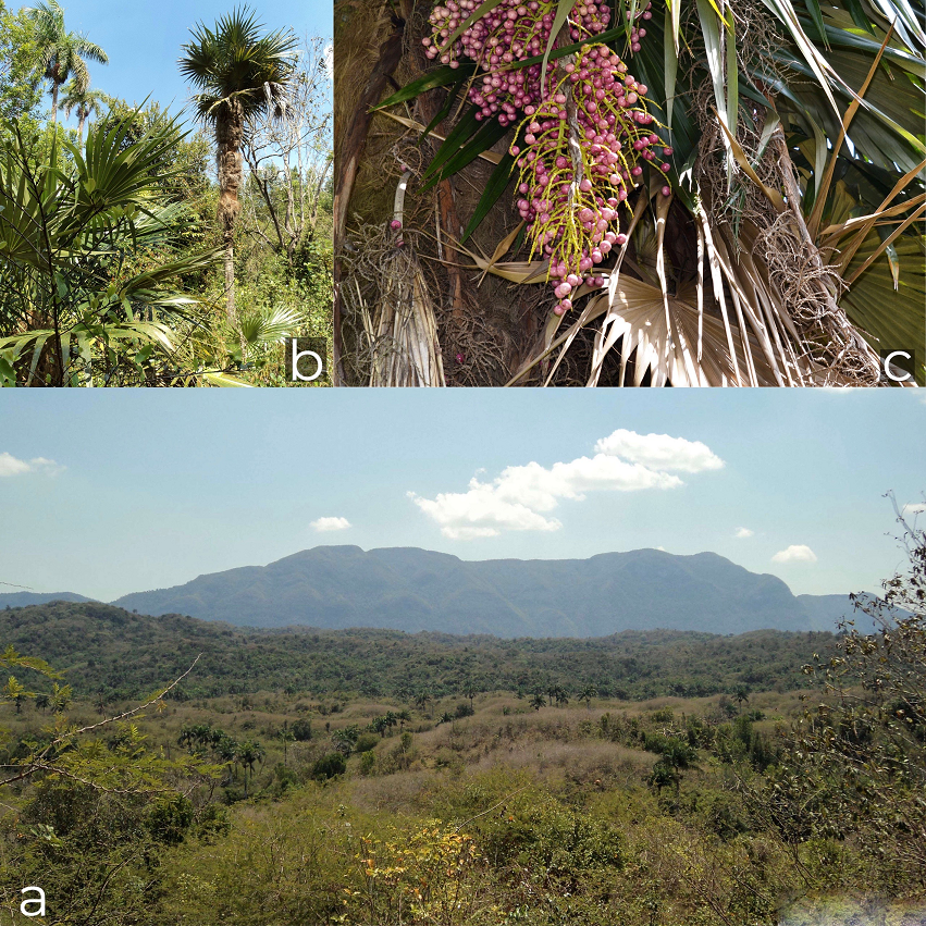
The study was conducted near Las Pozas (Fig. 1), a rural community located at 22°50’ N, 83°17’ W, Artemisa province, western Cuba, 17 km from the municipality of Bahía Honda. This locality is in the Pan de Guajaibón foothills, the highest elevation of Sierra del Rosario. The site presents an altitudinal range of between 36-156 m, decreasing from south to north. The carbonated soil is derived from igneous serpentinite rocks (brown tropical soil) and, in some cases, with serpentinite outcrops. Soil type is lithic Calcic Cambisol according to FAO-Unesco (1975). There are several hills at the base of Pan de Guajaibón to the north (Pinares, 2004). The habitat of C. crinita is a thorny xerophyte scrub on serpentine with secondary shrub vegetation in the most humid portion nearest to the gallery forest (Verdecia & Barrios, 2015). Annual mean temperature and precipitation values are 24.9 oC and 1,307 mm, respectively, which is characteristic of the Cajalbana district according to Borhidi (1996).

Figure 1 Coccothrinax crinita in its natural habitat. a) Region of Las Pozas, Artemisa Province; b) adult specimen; c) mature fruits.
Coccothrinax crinita is a palm 8 to 10 m height, with a trunk of up to 20 cm in diameter. Its leaves are large, soft and almost orbicular, with a pod that dissolves into very long and fine cream-colored fibers, which completely cover the trunk of the specimens in the first 15 to 20 years. The rhizodermis is hard, suberified and with several layers of epidermis. The fruits are globose, pink to purple when ripe and the seeds have cracks in the surface, giving them a brain-like appearance (Leiva, 1999; Fig. 1). Adult C. crinita specimens are scarce and unevenly distributed. In an area of 500 m2, we randomly chose 10 palms and sampled the soil and roots of each tree at a distance of 15-50 cm from the main stem by extracting 3 soil sample monoliths of 10 cm × 10 cm, which were mixed in order to form a compound sample, and removing roots from a depth of 20 cm. The soil and roots were placed into plastic bags for immediate transportation to the laboratory, where the roots were thoroughly rinsed with tap water and stored in 70% ethanol at 4 °C until subsequent analysis (Brundrett et al., 1996).
The rhizodermis was introduced into lactoglycerol to be cleared and stained for assessment of mycorrhizal colonization using the Phillips and Hayman (1970) technique, and the percentage of AMF colonization was quantitatively estimated following McGonigle et al. (1990). The process of clearing and staining caused deterioration of the rhizodermis, and it was therefore difficult to observe the fungal structures (arbuscules, vesicles, hyphal tangles, etc.) in detail for quantification. It was possible, however, to observe the presence of the fungus within the roots. For each sample, roots were cut into 1 cm pieces, and 30 randomly selected segments were mounted on slides with polyvinyl-lacto-glycerol (PVLG). Roots were scored under a microscope (CARL ZEISS model AXIOSKOP 2 Plus), at magnifications of 100-400×, for the presence of the following mycorrhizal structures: arbuscules, vesicles, intraradical and extraradical hyphae. Observations made under the microscope allowed identification of fragments of the blue-stained fungal structures. The presence of non-AMF fungal endophytes was also recorded, and these were categorized into 3 main groups: dark septate endophytes (DSE), thin external blue hyphae, and Rhizoctonia-like sclerotia.
Spores of the arbuscular mycorrhizal fungi present in the rhizosphere samples were extracted from the soil by wet sieving and decanting (Gerdemann & Nicolson, 1963). Briefly, 100 g air-dried soil samples were sieved (through 1,000, 140 and 40 μm mesh) followed by a density gradient centrifugation method (Oehl et al., 2003). The spores were mounted on slides with polyvinyl-lactic acid-glycerol (PVLG) (Koske & Tessier, 1983) or a mixture (1:1; v/v) of PLVG with Melzer’s reagent and viewed under a dissecting microscope (Brundrett et al., 1994). Subsequently, they were examined under a compound microscope. Identification was based on current species descriptions and identification manuals (International Culture Collection of Arbuscular and Vesicular-Arbuscular Endomycorrhizal Fungi at http://www.invam.caf.wvu.edu/Myc_Info/Taxonomy/species.html; Schenck & Pérez, 1990) and Blaszkowski´s web page (www.agro.ar.szczecin.pl/~jblaszkowski).
Results
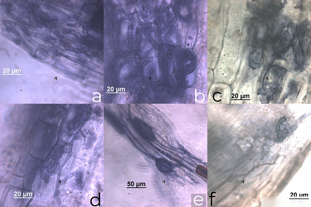
The C. crinita roots were colonized by AM fungi with a colonization average 60 ± 4.9%, being the highest root colonization 67% and the lowest 51% (Table 1). According to the presence and distribution of AMF structures in the root cortex, we propose an Arum-Paris type colonization pattern, according to Dickson (2004). Appressoria were formed by hyphae directly invading the epidermal cell with no apparent external modification. Internal arbuscules, vesicles as hyphal coils and dark septate endophytes were observed. We also observed thick extraradical hyphae running longitudinally, with some septa. The hyphae tended to form a coil within the first 3 adjacent epidermal cells and coils or loops within the adjacent exodermal and hypodermal cells (Fig. 2).
Table 1 Total root colonization in Coccothrinax crinita and fungal structures: arbuscules (A), vesicles (V), hyphal coils (HC), dark septate endophytes (DSE) and spore density in the rhizospheric soil.
| Plants | Total root colonization (%) | A | V | HC | DSE | Spore density (100 g soil) |
|---|---|---|---|---|---|---|
| 1 | 60 | x | x | x | x | 533 |
| 2 | 56 | x | x | x | 640 | |
| 3 | 59 | x | x | x | 980 | |
| 4 | 63 | x | x | x | 755 | |
| 5 | 65 | x | x | x | 1,023 | |
| 6 | 55 | x | x | x | 516 | |
| 7 | 64 | x | x | x | x | 669 |
| 8 | 67 | x | x | x | 1,112 | |
| 9 | 51 | x | x | x | 498 | |
| 10 | 60 | x | x | x | 832 | |
| Mean | 60 | 756 | ||||
| S.D. | 4.97 | 223.33 |

Figure 2 Arbuscular mycorrhizal root colonization. a) Longitudinal hyphae; b) thick longitudinal hyphae with tendency to coil in cortex cells; c) coils; d) vesicles; e) arbuscules; f) dark septate endophytes.
The hyphae traveled mainly in the longitudinal intercellular spaces of the inner cortical parenchyma. Coils invaded the adjacent cortical cells positioned both longitudinally and transversely to the root axis. AM fungi were never observed in the stele or endodermal cells. In old roots, hyphae (commonly septate) entered the cells of the inner cortex adjacent to the endodermis but did not penetrate the endodermal cells. Typical vesicles inter- and intracellular, elliptical or in lemon form, were observed. We also observed arbuscules, some of which had senescent aspect, and other fungi identified as dark septate endophytes (Fig. 2).
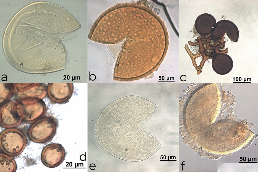
The average spore density in the rhizospheric soil was 756 ± 223.33 spores 100 g soil-1, ranging from 498 to 1,112 spores 100 g soil-1, equivalent to 5-11 spores per gram. We identified 16 morphologically distinct AMF species and/or morphospecies among the 10 samples studied (Fig. 3). More than half (7) belonged to the genus Glomus Tul and C. Tul, 2 species belonged to Funneliformis C. Walker & Schüßler, and 1 to Viscospora (T.H Nicolson) Sieverd., Oehl & G.A. Silva in the Glomerales. Three species belonged to Acaulospora Gerd. and Trappe and 1 to Kuklospora Oehl & Sieverd. in the Acaulosporales, while 1 belonged to Gigaspora Gerd. and Trappe and 1 to Scutellospora C. Walker and F.E. Sanders in the Gigasporales. Six AMF species could not be identified (Acaulospora sp. 1, Glomus sp. 1 “yellowish-brown in aggregates”, Glomus sp. 2 “brown small”, Glomus sp. 3 “reddish brown thick hyphae”, Glomus sp. 4 “big red in aggregates”, and Scutellospora sp. 1 “yellow light”). The most abundant AMF species were A. scrobiculata, G. clavisporum, G. glomerulatum and Glomus sp. 1 “yellowish-brown in aggregates”. These were found among 60-70% of all of the studied specimens. Spores of A. foveata, F. halonatum, F. mosseae and Glomus sp. 2 “brown small” were also relatively abundant, but with values of between 40 and 50% (Table 2; Fig. 3).

Figure 3 Some species of arbuscular mycorrhizal fungi. a) Scutellospora sp. “small yellow light”; b) Acaulospora foveata; c) Glomus sp. 3 “reddish brown thick hyphae”; d) Glomus “yellowish-brown in aggregates”; e) Acaulospora scrobiculata; f) Glomus halonatum cf.
Table 2 Presence-absence of arbuscular mycorrhizal fungal species in rhizospheric soil of Coccothrinax crinita.
| AMF species | Tree examined | Frequency (%) | |||||||||
|---|---|---|---|---|---|---|---|---|---|---|---|
| 1 | 2 | 3 | 4 | 6 | 6 | 7 | 8 | 9 | 10 | ||
| Acaulospora foveata Trappe & Janos | 1 | 0 | 1 | 1 | 0 | 0 | 1 | 0 | 1 | 0 | 50 |
| A. scrobiculata Trappe | 1 | 1 | 0 | 0 | 1 | 0 | 1 | 1 | 0 | 1 | 60 |
| Acaulospora sp. 1 “reddish brown” | 0 | 1 | 1 | 0 | 0 | 1 | 0 | 0 | 0 | 0 | 30 |
| Funneliformis halonatum (S.L. Rose & Trappe) Oehl, G.A. Silva & Sieverd | 0 | 0 | 1 | 0 | 0 | 1 | 0 | 0 | 1 | 1 | 40 |
| Funneliformis mosseae (T.H. Nicolson & Gerd.) C. Walker & A. Schüssler | 0 | 1 | 0 | 0 | 1 | 1 | 1 | 0 | 0 | 1 | 50 |
| Glomus clavisporum (Trappe) Almeida & Schenck | 1 | 1 | 0 | 1 | 1 | 0 | 1 | 1 | 1 | 0 | 70 |
| Glomus glomerulatum Sieverd | 1 | 1 | 0 | 1 | 0 | 1 | 0 | 1 | 1 | 1 | 70 |
| Glomus tortuosum Schenck & Smith | 0 | 0 | 0 | 0 | 1 | 0 | 0 | 1 | 0 | 0 | 20 |
| Glomus sp. 1 “yellowish-brown in aggregates” | 1 | 0 | 1 | 0 | 1 | 1 | 1 | 0 | 1 | 1 | 70 |
| Glomus sp. 2 “brown small” | 0 | 0 | 1 | 1 | 0 | 0 | 0 | 1 | 1 | 0 | 40 |
| Glomus sp. 3 “reddish brown thick hyphae” | 0 | 1 | 0 | 0 | 1 | 0 | 0 | 0 | 0 | 0 | 20 |
| Glomus sp. 4 “big red in aggregates” | 0 | 0 | 0 | 1 | 1 | 0 | 0 | 0 | 0 | 0 | 20 |
| Viscospora viscosa (T.H Nicolson) Sieverd., Oehl & G.A. Silva | 1 | 0 | 0 | 0 | 0 | 0 | 1 | 0 | 1 | 0 | 30 |
| Kuklospora kentinensis (Wu & Liu) Oehl & Sieverd. | 0 | 1 | 0 | 0 | 0 | 0 | 1 | 0 | 0 | 1 | 30 |
| Gigaspora decipiens Hall & Abbott | 0 | 0 | 1 | 0 | 0 | 1 | 0 | 0 | 0 | 1 | 30 |
| Scutellospora sp. “small yellow light” | 1 | 0 | 0 | 1 | 0 | 0 | 0 | 0 | 1 | 0 | 30 |
Discussion
The presence of AMF colonization in C. crinita roots and spores in its rhizospheric soil is important for the potential conservation of this species reported as Critically Endangered in the Flora of Cuba Red List (González-Torres et al., 2016). The species probably presents ecological specificity and its AMF could be a vital element in its survival and maintenance (McGonigle & Fitter, 1990).
Al-Yahya’ei (2008) and Fisher and Jayachandran (1999) discussed the difficulty of distinguishing AMF structures in palm roots. However, in this study, intraradical hyphae, vesicles and arbuscules were observed (Fig. 2), demonstrating the arbuscular mycorrhizal status of C. crinita. Usually the presence of arbuscules is considered the best indicator of function in the AM association (Corkidi & Rincón, 1997; Gange & Ayres, 1999); however, it has been recognized that arbuscules are not the only structures for nutrient translocation, and the vesicles and intraradical hyphae are indicators of current and past colonization (Allen, 1991). In this study, the arbuscules in the roots of C. crinita were not abundant, indicating that all fungal structures contribute to the host-fungi symbiosis (Smith & Smith, 1997). Arbuscular mycorrhizal fungal root colonization in C. crinita was higher than 60%, a value similar to that found for Astrocaryum mexicanum Liebm in tropical rainforest (Núñez-Castillo & Álvarez-Sánchez, 2003) and Thrinax radiata Lodd. ex Schult. in sand dunes (Polanco et al. 2013). On the other hand, colonization was lower in other Arecaceae, such as C. readii (27.7% during the rainy season, Polanco et al., 2013), D. orthacanthos (33% in mature tropical dry forest, Ramos-Zapata et al., 2006a) and peach palm (Bactris gasipaes Kunth) cultivated in the Amazon, from 13.54 - 43.95% (da Silva Júnior & Cardoso, 2006).
We classified the colonization pattern in C. crinita as Arum-Paris type, arbuscules with a certain degree of degradation were distinguished (Fig. 2c, d) and hyphal coils were also observed in the roots (Fig. 2a, b). As has been observed in Areca catechu L., Borassus flabellifer L. and Nypa fruticans Wurmb (Sengupta & Chaudhuri, 2002), Bactris gasipaes Kunth (da Silva Júnior & Cardoso, 2006), S. repens (Fisher & Jayachandran, 1999) and Phoenix paludosa Roxb, which is defined as a combination of “both types” according to Smith & Smith (1997).
Mycorrhizal symbiosis ranges from facultative to obligate, depending on the successional stage, seed size and nutrient requirements (Allen et al., 2003; Huante et al., 1993; Sharma et al., 2001; Siqueira & Saggin-Junior, 2001; Siqueira et al., 1998). Species such as D. orthacanthos were considered as obligate mycorrhizal in the semi-evergreen tropical forest of Quintana Roo, Mexico according to Ramos-Zapata et al. (2006a), while C. readii was considered facultative to the association with AMF fungi, as proposed by Polanco et al. (2013), since he found a low proportion of arbuscules and spore density in the soil. In our case, some experiments are necessary in order to establish the mycorrhizal dependency status of that palm. Plants with some degree of mycorrhizal dependence can increase the concentration of phosphorus in their tissues, while their seedlings have a greater survival capacity due to the hyphal network established in the soil, which is very important for a critically endangered species (Grime et al., 1987; Francis & Read, 1995).
The richness and density of spores, as well as mycorrhizal colonization, are important variables to infer the ability of the soil to produce mycorrhizae and to assess soil health (Cuenca, 2015). The spore density in rhizospheric soil of C. crinita was higher than that of the Amazon palms Attalea speciosa Mart (100 to 302 spores 100 g-1 dry soil) (Nobre et al., 2018) and B. gasipaes (54 to 96 spores 100 g-1 dry soil) (da Silva Júnior & Cardoso, 2006). On the other hand, the density was lower than that of E. guineensis (1,289-1,741 spores 100 g-1 dry soil) in coastal and Amazonian zones of Ecuador (Maldonado et al., 2008), S. repens (260-2,200 spores 100 g-1 dry soil) from the southeastern USA (Fisher & Jayachandran, 1999) and D. orthacanthos (250 and 350 spores 100 g-1 dry soil) from Quintana Roo, Mexico (Ramos-Zapata et al., 2006a).
Coccothrinax crinita has a high AMF richness similar to that reported by Nobre et al. (2018) in A. speciosa under natural conditions, but lower than that found in palm plantations in Oman (25 species) (Al-Yahya’ei et al., 2011). Glomus was the most frequently found genus in the C. crinita rhizosphere, as was observed in other palms (Fisher & Jayachandran, 1999; Guevara & López, 2007; Al-Yahya’ei et al., 2011).
Acaulospora has also been found in palms in the Brazilian Amazonia (Nobre et al., 2018; Silva et al., 2006; Trufem et al., 1994). Consequently, some Glomus and Acaulospora species could be considered as generalist AM fungi, although further studies would be necessary in order to confirm this specialist status.
The present study has shown the association of this palm with AMF in their natural habitat and, in the near future, some experiments inoculating these fungi could provide some tools to help improve the conservative management of C. crinita. An understanding of the mycorrhizal relationships of C. crinita, as well as the AMF species associated with this palm under natural conditions, could contribute to its conservation and given its multiple uses, enable cultivation for economic purposes.


 nueva página del texto (beta)


