Introduction
Arrhythmias are uncommon in pediatrics. However, they occur more frequently during the first days of life due to the period of relative electrical instability1. They are more frequent in preterm newborns, with episodes of sinus bradycardia and junctional escape rhythms, and do not usually require treatment. Acquisition of an electrocardiogram (ECG) during the episode is crucial for diagnosis2.
Atrial flutter has a low incidence in children. It is characterized by a rapid atrial rate of approximately 300 beats per minute (bpm) or more and distinctive sawtooth P waves, called F waves3.
Although atrioventricular conduction can be variable, the most common is 2:1 conduction, which is usually well-tolerated; however, 20% of cases may present heart failure4. In adults and older children, flutter is generally associated with structural heart disease, one of the complications that can appear after surgical correction of different congenital heart diseases. A possible etiology should be sought in the first episode, ruling out cardiac dysfunction or associated heart disease. The evolution is usually satisfactory if the arrhythmia is not present and begins in the neonatal period5. Although the evolution of this arrhythmia in the neonatal period is benign, it requires intensive treatment with drugs or electrical cardioversion at the onset time. The recommended treatment for both stable and unstable neonates is synchronized electrical cardioversion. As a second choice, the recommended drugs are digoxin and flecainide or amiodarone if there is no therapeutic effect with the first two agents. According to the literature reviewed, cardioversion with direct current appears to be the most effective treatment for atrial flutter6.
Due to the low frequency of this disease in pediatric patients without structural heart disease, we present a retrospective review of the clinical characteristics of the cases under follow-up in our center. Most showed a benign evolution of the arrhythmia since those diagnosed in the neonatal period did not present new episodes of atrial flutter after treatment with electrical cardioversion. However, those diagnosed at a later stage (5-7 years of age) required electrical ablation of the ectopic focus due to failure of first-line treatments.
Clinical cases
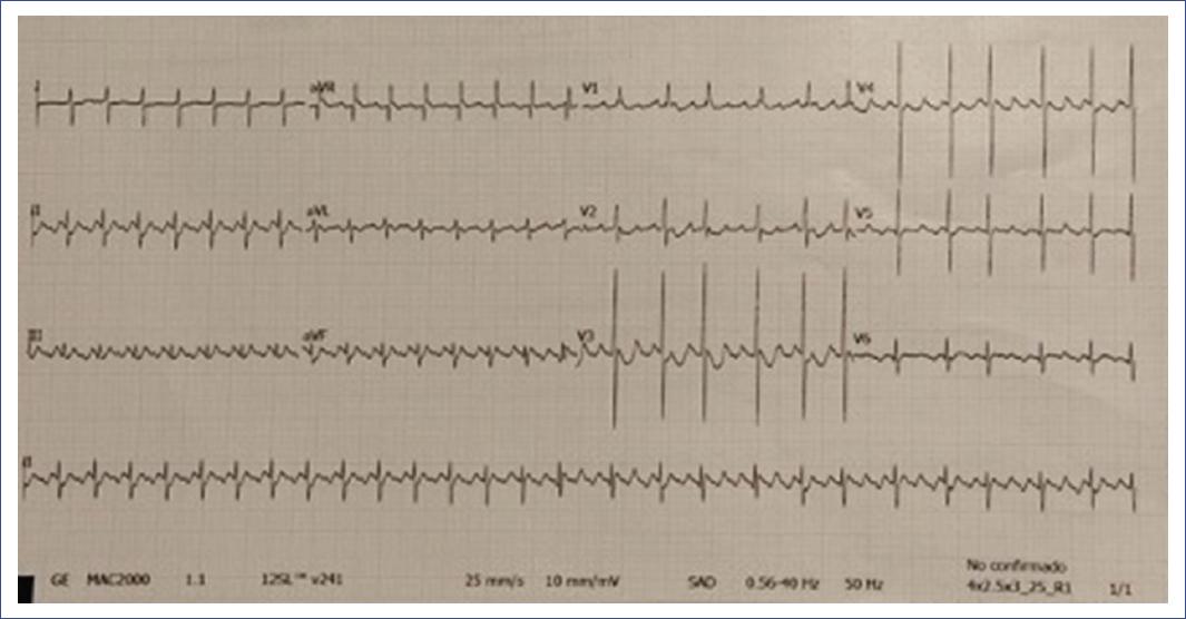
We conducted a retrospective review of all cases of atrial flutter in children (0-15 years) in the last six years (2015-2021) at a tertiary pediatric hospital. After parental authorization and consent, an analysis was performed by reviewing patients’ medical records. Seven cases were reviewed: six males and one female, five diagnosed in the perinatal period, and the other two at school age. In three of them, tachycardia was already evident in the fetal period; in the rest, pregnancy control and prenatal ultrasound had been normal, with flutter appearing later (Figure 1). In all cases, structural heart disease was excluded by echocardiography.

Figure 1 Electrocardiogram of a newborn with tachycardia at 200 bpm and sawtooth F waves compatible with atrial flutter with 2:1 and 3:1 block.
The findings of each case studied are described below.
Case 1
We describe the case of a male neonate who, during cardiac screening performed in our hospital to measure preductal and postductal oxygen saturation between the first 6 and 24 hours of life and heart rate, showed tachycardia with a frequency of 200 bpm, although he was asymptomatic. An ECG was performed, and atrial flutter was diagnosed at 20 hours of life. Electrical cardioversion was performed, and the patient did not present new episodes. Clinical evolution was good, with sinus rhythm at 180 bpm, and he was discharged five days after birth. He showed no other arrhythmias during follow-up with his pediatrician at annual check-ups.
Case 2
This case is a preterm twin male neonate who, at birth, required respiratory support. He was transferred to the Neonatal Intensive Care Unit (NICU) and, once monitored, presented an episode of flutter at 36 hours of life, which was resolved with electrical cardioversion with no clinical complications; at the same time, respiratory distress was resolved. The patient presented two more episodes of atrioventricular reentrant tachycardia mediated by an occult accessory pathway 10 and 17 days after birth, requiring flecainide during the first months of life. During his subsequent outpatient follow-up in pediatric cardiology consultations, he did not present new episodes of arrhythmias and was discharged at 2 years of age.
Case 3
The third case is a male neonate born by emergency cesarean section due to fetal tachycardia at 37+2 weeks of gestation, with a birth heart rate of 230 bpm. The ECG showed sawtooth F waves characteristic of flutter, with a 2:1 ventricular block confirmed after administration of intravenous adenosine at 0.25 mg/kg body weight. Electrical cardioversion was performed with a 2J charge at birth, and second cardioversion was required with a 3J shock, which was effective. The patient did not present arrhythmias again and, therefore, did not require medical treatment. He was discharged at one month of life, living an everyday life without any pharmacological treatment. At his last pediatric cardiology control appointment, at four years of age, his electrocardiogram showed sinus rhythm with normal axes, voltages, and repolarization, for which he was discharged from the service.
Case 4
This case is a female preterm neonate diagnosed with fetal supraventricular tachycardia who received prenatal treatment with flecainide and digoxin. Labor was induced at 34+2 weeks of gestation due to persistent fetal tachycardia despite prenatal treatment. At birth, the neonate presented tachycardia > 200 bpm, with ineffective respiratory effort, requiring respiratory support. Up to three electrical cardioversions and treatment with flecainide were necessary until the patient was four months old. Treatment was discontinued without the appearance of new arrhythmias. The patient was discharged from cardiology at four years of age; she has not presented new episodes, and her electrocardiograms showed no alterations.
Case 5
This case of a male neonate with a prenatal diagnosis of fetal flutter and treatment with digoxin during pregnancy; at birth, he presented sinus bradycardia that resolved spontaneously without requiring any treatment. There were no postnatal flutter episodes.
Case 6
This case corresponds to a 5-year-old male patient with a family history of Brugada syndrome who was referred to the hospital for suspected Brugada syndrome after an abrupt episode of loss of consciousness. He presented with an ECG compatible with Brugada syndrome type 1, showing right bundle branch block and dolphin fin descending ST elevation in the right precordial leads and V2 with negative T wave. The patient received an implantable cardioverter-defibrillator (ICD) at 5 years of age. During follow-up, an episode of atrial flutter with a rate of 200 bpm with 2:1 block was detected at 7 years of age in the Holter recording of the ICD without clinical correlation. The original focus was electrically suppressed without incident. The patient did not present any more tachyarrhythmias in his subsequent evolution.
Case 7
This last case corresponds to an eleven-year-old male patient who came to the emergency department for an episode of syncope. His family history included the sudden death of his 26-year-old mother, the exact cause being unknown. An ECG was performed in the emergency room, showing an episode of polymorphic ventricular tachycardia with a rate of 260 bpm. A subcutaneous Holter was implanted, showing how this tachycardia triggered an episode of torsade de pointes lasting 1 minute, with spontaneous recovery after five seconds of asystole accompanied by another syncope. A genetic study was performed, and the patient was diagnosed with polymorphic ventricular catecholaminergic tachycardia with a mutation in the ryanodine gene (RYR2 14311G>A). Treatment with bisoprolol and flecainide was started, and left cervical sympathectomy was performed due to the recurrence of tachycardias. During follow-up, different episodes of arrhythmias were detected in the implanted subcutaneous Holter monitor, including atrial flutter, whose ectopic foci were subsequently suppressed with no subsequent incidents.
The clinical characteristics, treatment, and evolution of the patients are summarized in Table 1.
Table 1 Clinical characteristics, treatment, and evolution of patients
| Gender | Age at diagnosis | Prenatal diagnosis | Prematurity | ECG | Treatment | Relapses | Other associated diseases | |
|---|---|---|---|---|---|---|---|---|
| Case 1 (Figure 1) | Male | Neonate (20 hours of life) | No | Yes (35 GW) | Flutter with 2:1 block | Electrical cardioversion | No | No |
| Case 2 (Figure 2) | Male | Neonate (36 hours of life) | No | Yes (30+1 GW) | Flutter | Electrical cardioversion + flecainide in the first months of life | Two episodes of AV reentrant tachycardia mediated by hidden accessory pathway at 10 and 17 days of life | No The genetic study for the SCN5A gene was negative |
| Case 3 | Male | Neonate (at birth) | Yes Urgent cesarean section due to fetal tachycardia |
No | Flutter at 250 bpm | Electrical cardioversion | No | No |
| Case 4 | Female | Neonate | Yes Fetal SV tachycardia Prenatal digoxin and flecainide |
Yes (34+2 GW) | Flutter > 200 bpm | Electrical cardioversion x3 + flecainide up to 4 months of life | No | No |
| Case 5 | Male | Neonate | Yes Fetal flutter on prenatal ultrasound Prenatal digoxin |
No | At birth sinus bradycardia at 80 bpm | Not required | No | No |
| Case 6 | Male | 5 years | No | No | ECG compatible with type 1 Brugada syndrome
At 7 years of age, Holter monitoring showed an episode of flutter at 200 bpm with a 2:1 block |
Ablation of the original flutter focus by catheter two years after diagnosis | No | Type 1 Brugada syndrome |
| Case 7 | Male | 11 years | No | No | Flutter, polymorphic ventricular tachycardia, atrial tachycardia, atrial fibrillation | Ablation of the ectopic foci by catheter | Several subsequent episodes of arrhythmias | With different types of arrhythmias in successive Holter recordings |
AV, atrioventricular; bpm, beats per minute; ECG, electrocardiogram; GW, gestation weeks; SV, supraventricular.
The outcome of all patients diagnosed in the pre-or neonatal period was satisfactory. However, when the episodes occurred later (5 and 7 years of age), they were associated with channelopathies (Brugada syndrome and catecholaminergic polymorphic ventricular tachycardia), with necessary ablation due to poor response to pharmacological treatment (Figure 2).
Discussion
Flutter is an example of macro reentrant atrial tachycardia; in 90% of cases, the right atrium is the location of the original focus of the reentrant circuit6. The incidence of this disease is extremely low in children. The incidence of atrial flutter in pediatrics is estimated at 0.01%, which is an underestimated figure due to the low rate of clinical suspicion7. Our series demonstrates the low incidence of atrial flutter in the pediatric population (0.003%), with only seven cases over 6 years in a reference population of almost 200,000 subjects aged 0-14 years.
This review also intended to differentiate between two very distinct groups within the same pathology: cases in newborns and those diagnosed later in life. We can define evident clinicopathological characteristics when flutter occurs in the neonatal period, as in the first five cases, since most appear in structurally normal hearts, present a benign course with an adequate response to electrical cardioversion, and a tendency to spontaneous resolution over time8,9. The evolution of the cases diagnosed in the neonatal stage is considered benign since electrical cardioversion was effective in all the cases studied (as we have also found in the literature reviewed), with no patients experiencing tachyarrhythmias or heart pathology during follow-up in the subsequent years9. Case 3 required two electric shocks, and case 4 required cardioversion on three occasions; this approach is not considered a factor of poor prognosis or malignancy since it resolved without incident, and the patients remain asymptomatic.
Yılmaz-Semerci et al. described in 2018 that flutter or atrial flutter in the newborn occurs during the first 7 days of life, as we observed in our neonatal cases, in whom it occurred at birth, at 20 and 36 hours after birth or prenatally10. However, when an older child or adolescent is diagnosed with atrial flutter, the prognosis worsens, and the probability of concomitant associated heart disease is more significant, as well as presenting other electrical disorders such as Wolf-Parkinson-White syndrome or channelopathies. Consistent with what Nieto et al. described in their 2020 publication, treatment is less effective in older patients, as in our cases, at 5 and 7 years of age. These patients require much closer follow-up for the rest of their lives, as they are more likely to have other associated heart diseases11.
Although atrial flutter is a rare condition in pediatrics and is not usually associated with structural heart disease, it must be diagnosed and treated effectively. The primary postnatal treatment is electrical cardioversion, aiming at achieving sinus rhythm.
The reviewed literature indicated that neonatal recurrence is rare and does not usually require subsequent treatment, as in this review of cases in our hospital9,11. However, flutter in older patients without congenital heart disease is generally associated with channelopathies, presenting a worse evolution and greater refractoriness to medical treatment, as in our last two cases.
In conclusion, it is essential to emphasize the difference in the evolution of patients diagnosed with flutter in our hospital according to their age at the time of diagnosis. According to our experience, cases of neonatal flutter evolved satisfactorily with electrical cardioversion. Furthermore, in one of the cases presented, cardioversion was not necessary since, after birth, the patient did not show fetal tachycardia detected prenatally. Moreover, no underlying heart disease was found in either fetal or neonatal cases. Regarding the two patients diagnosed at an older age (5-7 years), we highlighted the presence of associated heart disease. One had Brugada syndrome, and the other had catecholaminergic tachycardia with a genetic mutation, conditions that confer patients a worse prognosis and require more invasive treatments and closer follow-up.


