INTRODUCTION
Coralline algae are abundant and ecologically important aquatic vegetation in coastal areas around the world (Foster 2001). These algae establish large, dense beds in several regions of the world, including the Gulf of California, and dense mats in rocky shores in the North Pacific and North Atlantic, forming important micro- and macrohabitats for invertebrates and algae (Foster 2001, Steller et al. 2003). Furthermore, coralline algae are a major component of the diet or settling substrata of commercially important fishery resources such as lobsters and abalone (Jernakoff et al. 1993, Linnane et al. 2000). Due to their abundance and ability to calcify, coralline algae are recognized as key components of carbon cycles in shallow coastal waters (Chisholm 2000, Martin et al. 2006). The calcium carbonate skeleton of these algae serves as a physical defense and plays an important role in their light absorption (Vásquez-Elizondo and Enríquez 2017), but it also makes coralline algae vulnerable to ocean acidification (Díaz-Pulido et al. 2012). An increasing body of evidence indicates that thermal stress severely affects coralline algae performance (Martin and Gattuso 2009, Vásquez-Elizondo and Enríquez 2017).
Photosynthetic rates of coralline algae have been generally investigated using gas-exchange procedures. Pulse amplitude modulated (PAM) fluorometry represents a clear advantage to traditional methods due to its versatility, nonintrusive nature, and rapid evaluation of photosynthetic activity (Maxwell and Johnson 2000). In PAM fluorometry, chlorophyll a fluorescence associated with photosystem II (PSII) is used to assess primary reactions and quenching mechanisms of the photosynthetic apparatus (Maxwell and Johnson 2000, Longstaff et al. 2002, Beer and Axelsson 2004). In addition, the electron transport rate (ETR) from PSII to photosystem I (PSI) can be calculated and used as a proxy for photosynthesis, but its effectiveness depends on the linearity between ETR and the carbon dioxide-oxygen evolution (Franklin and Badger 2001, Beer and Axelsson 2004).
In land vegetation, especially in C4 plants, ETR and carbon uptake generally maintain a linear relationship (Edwards and Baker 1993), while in marine macrophytes, the relationship between gross photosynthesis (GPS) and ETR is generally species-specific and varies as a function of light acclimation, temperature, and nitrogen availability (Cabello-Pasini et al. 2000, Figueroa et al. 2003, Cabello-Pasini and Figueroa 2005). For example, the GPS vs. ETR relationship is linear in Ulva rigida and Macrocystis pyrifera, but it deviates at high irradiance in Ulva fasciata (Carr and Bjork 2003, Colombo‐Pallotta et al. 2006). Only one study demonstrated a linear association between the GPS and the relative ETR of the rhodolith Phymatolithon lusitanicum (Sordo et al. 2020). Therefore, extensive research is needed to evaluate the utility of the ETR in coralline algae.
The fixation of carbon dioxide in marine macrophytes is mainly dependent on irradiance, temperature, and nutrient levels (Falkowski and Raven 2007). In coastal shallow environments, marine macrophytes are exposed to daily and seasonal fluctuations in temperature, which induce variation in photosynthetic rates, as well as pigment, protein, and fiber contents (Cabello-Pasini et al. 2003, 2004). As a consequence, algae exposed to fluctuating conditions are expected to suffer less from the adverse effects of environmental variation (Martone et al. 2010, Williamson et al. 2017). The temperature effect on coralline algae physiology is well studied, but little is known about this effect on the ETR. Short-term increases in natural temperature regimes increase photosynthetic rates, but this varies among species, light acclimation and exposure times (Guy-Haim et al. 2016, Vásquez-Elizondo and Enríquez 2016), whereas thermal stress typically induces a loss in performance (Martin and Gattuso 2009, Webster et al. 2011).
A detailed description of the photosynthetic response to temperature gradients will increase our ability to understand different response mechanisms and to interpret more complex and diverse scenarios. The ability to rapidly evaluate the photosynthetic response (i.e., ETR) can benefit future research after the limitations of this approach are defined. Consequently, the objective of this study was to simultaneously evaluate the short-term effect of temperature (10-35 °C) on the photosynthesis, respiration, and ETR of 3 articulated coralline algae from the Pacific North Coast of Mexico while evaluating the relationship between the ETR and GPS.
MATERIALS AND METHODS
Biological material
Samples of the articulated coralline algae Lithothrix aspergillum, Corallina officinalis, and Bossiella orbigniana were collected in the intertidal zone of Punta Morro, Ensenada, Baja California, Mexico, in March 2006. Seawater temperature was between 17 and 20 °C, and the surface irradiance reached 1,000-1,200 µmol quanta·m-2·s-1 at noon on sunny days. The collected material was transported to the nearby laboratory of the Autonomous University of Baja California (UABC). Samples were cleaned of epiphytes, rinsed, and placed in 20-L containers with filtered seawater (0.45 µm) at 20 °C, with constant aeration to promote water circulation, and at 80 µmol quanta·m-2·s-1. The water was changed every 12 h. All experiments were conducted within 2 days after sample collection.
Oxygen evolution and chlorophyll a fluorescence
Simultaneous measurements of oxygen and chlorophyll a fluorescence were performed on the algae thalli. Oxygenic photosynthetic rates were determined using polarographically measured rates of steady-state oxygen evolution on apical thalli segments (n = 4, ≈0.05-0.10 g fresh weight). All measurements were conducted in 5-mL jacketed chambers (Rank brothers; Cambridge, UK) where temperature was controlled using a water-circulating bath (ISOTEMP 1016S, Fisher Scientific; Hampton, New Hampshire, USA; stability 0.5 °C). A preincubation period in darkness (0.5 h) at the experimental temperature was applied by placing the algae in 50-mL tubes filled with filtered seawater in the water-circulating bath system reservoir. Oxygen evolution was recorded for 8-10 min from darkness (respiration) through a series of increasing irradiances (0 to 960 µmol quanta·m-2·s-1) using neutral density filters (Lee filters; Osram, UK) placed in front of the light source (Quartzline 300 W). An experimental determination (a photosynthesis-irradiance curve of 0-960 µmol quanta·m-2·s-1) was performed in the same experimental segment to ensure that the response was due to the light history. The oxygen concentration was maintained between 20% and 40% by bubbling the water in the chambers with nitrogen (N2) at the beginning of the light curve, when no water exchange was used. Irradiance values within the chambers were determined using a miniature PAR quantum sensor (Diving PAM; Walz, Germany) previously calibrated against a cosine-corrected sensor (LI-190SA) connected to a portable radiometer (LI-1400, LI-COR; Lincoln, Nebraska, USA). The maximum photosynthesis (Pmax) was calculated from the average maximum values above saturating irradiances, and the initial slope of the photosynthesis vs. irradiance (P-E) curve (α) was determined using a least-square regression analysis for the values under saturating irradiance showing a linear response to irradiance of each independent curve. The saturating irradiance (Ek) was determined as the ratio between Pmax and α. The metabolic coefficient Q10 was calculated as the ratio of average metabolic activity at a given final temperature (T °C + 10) to the average metabolic activity at a given initial temperature as long as the response was linear.
In vivo chlorophyll fluorescence of PSII was determined (n = 4) using a portable PAM fluorometer (Diving PAM; Walz, Germany) simultaneously during oxygen evolution measurements by introducing miniature fiber optics (Diving F1, Walz) into the oxygen chambers and keeping it at a 45° angle from the thalli throughout the experiments. The 2-mm diameter fiber optics was translucent to prevent shading of the thalli by the fiber. Basal fluorescence (Fo) and maximum fluorescence (Fm) in dark-acclimated samples (0.5 h) were determined before and after applying a saturating actinic light pulse (>3,000 µmol quanta·m-2·s-1, 0.8 s), respectively. Variable fluorescence (Fv) was determined as the difference between Fm and Fo, and maximum photochemical efficiency (Fv/Fm) was calculated (Schreiber 2004). Similarly, the photochemical efficiency or effective quantum yield of PSII (∆F/Fmʹ) was determined in light-acclimated thalli at each experimental irradiance after light steady oxygen evolution. ETR was determined as follows:
where absorptance, APAR, is the fraction of incident light absorbed by the algal thalli between 400 and 700 nm, E is the irradiance reaching the thalli, and 0.15 is the fraction of APAR directed to PSII used for red macroalgae (Orzymski et al. 1997). Absorbance and transmission in the algae thalli was calculated as described in Vásquez-Elizondo et al. (2017). Additionally, reflectance in coralline algae is considerably high compared to other non-calcifying algae, resulting in an absorption between 45% and 85% of the incident light, depending on thalli thickness and pigmentation, and a standard value of 0.7 for APAR was used (median value, see Vásquez-Elizondo and Enríquez 2017). The maximum ETR values, the initial slope of the ETR vs. irradiance curve, and the saturation irradiance for the ETR (Ek ETR) were calculated as previously described for the oxygen evolution curves. The algal dry weight (DW) was determined on an analytical scale after drying the algae for 48 h at 60 °C.
Statistical analysis
Differences in photosynthetic parameters (GPS, ETR) as a function of temperature were evaluated using a one-way analysis of variance after testing for normality and homoscedasticity. All pairwise multiple comparisons were conducted using Tukey’s test. If necessary, nonparametric Kruskal-Wallis tests were conducted. The significance of the correlation between ETR and GPS was tested using Pearson’s correlations. The significance level was established at P < 0.05. All statistical analyses were conducted in JAMOVI.
RESULTS
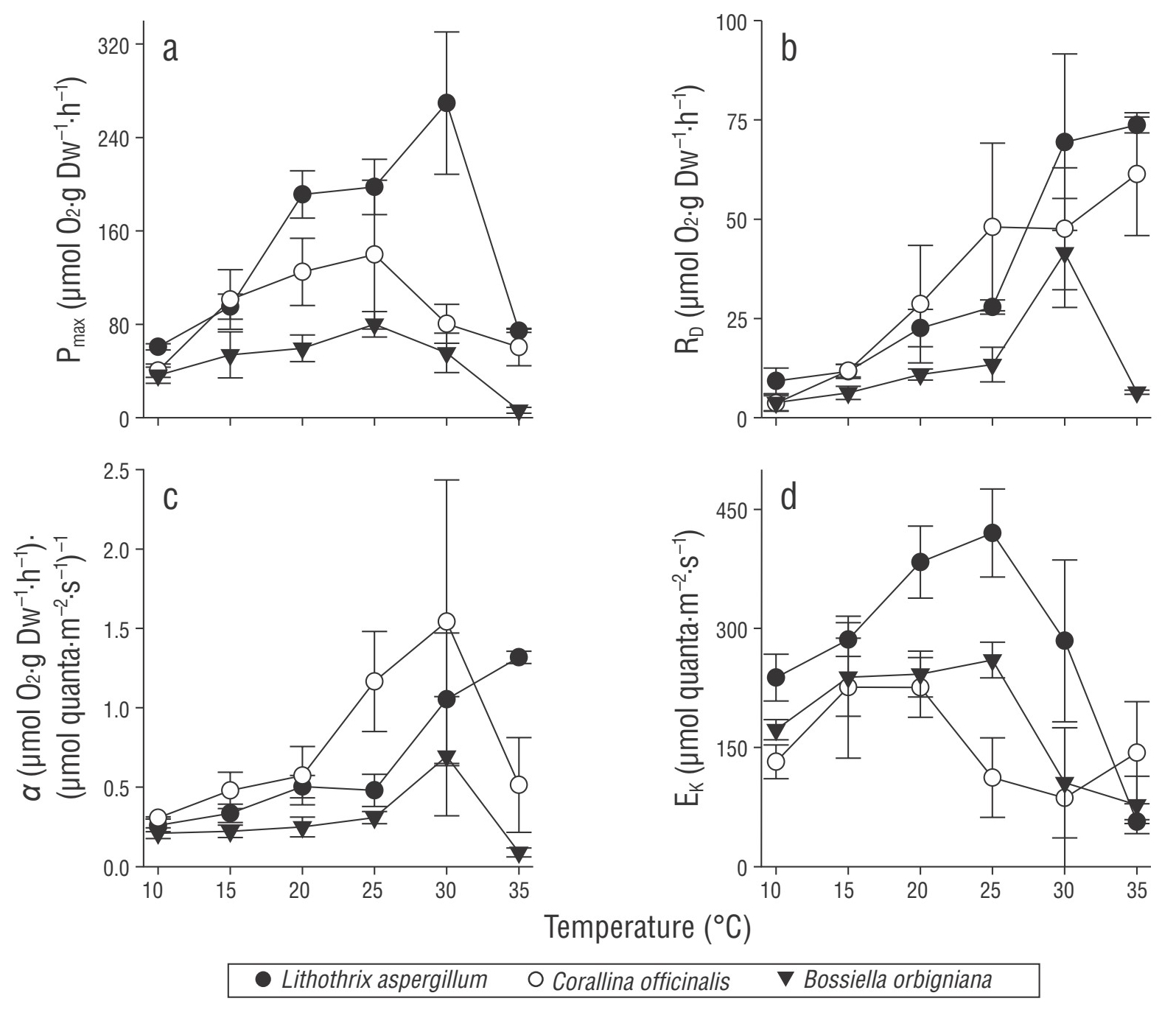
Temperature significantly affected the photosynthetic parameters calculated from the P-E curve (Fig. 1, S1) in all coralline algae studied (P < 0.05; Tables 1, S1). In general, Pmax, dark respiration (RD), α, and Ek increased linearly with increasing temperature to an optimum, followed by a strong decrease at higher temperatures (Fig. 1). The responses to temperature were species-specific, and the calculated metabolic rates showed distinct physiological thresholds. A linear increase in Pmax with increasing experimental temperature up to a maximum value was observed from 10 to 30 °C in L. aspergillum and from 10 °C to 20-25 °C in the remaining species (Fig. 1a). The maximum Pmax values were 269.3 (±60.8), 139.7 (±63.6), and 80.0 (±10.9) µmol O2·g DW-1·h-1 for L. aspergillum, C. officinalis, and B. orbigniana, respectively. After this maximum value, Pmax decreased substantially: 4-, 3-, and 12-fold for L. aspergillum, C. officinalis, and B. orbigniana, respectively. In general, RD (Fig. 1b) increased linearly over the full range of experimental temperatures in L. aspergillum and C. officinalis and reached a maximum at 30 °C in B. orbigniana (with a dramatic increase from 25 to 30 °C), followed by a strong decrease at 35 °C. The highest RD values were approximately 73.7 (±2.0), 61.3 (±15.4), and 41.5 (±13.7) µmol O2·g DW-1·h-1 for L. aspergillum, C. officinalis, and B. orbigniana, respectively. The initial slope of oxygen evolution (αoxy, Fig. 1c) showed a similar trend to Pmax and RD; it increased with increasing temperature to a maximum, from 10 to 30 °C in C. officinalis and B. orbigniana and at 35 °C in L. aspergillum (Fig. 2c). Ek showed a similar response among the species to increasing temperature and was generally higher for L. aspergillum, particularly between 20 and 25 °C (Fig. 1d). In C. officinalis, Ek increased in samples incubated from 10 to 20 °C, followed by a significant decrease and stabilization from 25 to 35 °C. In the remaining species, Ek increased from 10 °C to a maximum at 25 °C in B. orbigniana and 20 °C in L. aspergillum, followed by a strong decrease. The photosynthesis to respiration (P:R) ratios for all species were relatively high at lower temperatures (~7-13) but significantly decreased with increasing temperatures (30-35 °C) to values of approximately 1 (Tables 1, 2). The metabolic quotient Q10 was variable but generally decreased with increasing temperature in all species (Table 2). While L. aspergillum and C. officinalis showed the greatest photosynthetic Q10, C. officinalis and B. orbigniana showed the greatest respiratory Q10.
Table 1 One-way analysis of variance/Kruskal-Wallis results for the comparison between the photosynthetic descriptors (oxygen/chlorophyll fluorescence) and temperature in the studied coralline algae. Abbreviations: Pmax, maximum photosynthesis rate; RD, dark respiration; α, initial slope of the photosynthesis vs. irradiance curve; Ek, saturation irradiance; P:R, photosynthesis to respiration ratio; Fv/Fm, maximum photochemical efficiency; ETRmax, maximum electron transport rate; αETR, initial slope of the ETR vs. irradiance curve; and Ek ETR, saturation irradiance for the ETR. All analyses were conducted among the 5 temperature treatments except in Bossiella orbigniana (n = 4 treatments, no data for αETR and Ek ETR for 35 °C). See Table S1 for post hoc multiple comparisons.
| Oxygen evolution | Chlorophyll a fluorescence | ||||||||
| Species | Descriptor | F, H | d.f. | P | Descriptor | F, H | d.f. | P | |
| Lithothrix aspergillum | Pmax* | 21.10 | 5,18 | <0.001 | Fv/Fm * | 15.5 | 5.0, 18.0 | 0.008 | |
| RD* | 20.80 | 5,18 | <0.001 | ETRmax | 86.6 | 5.0, 7.0 | <0.001 | ||
| α* | 19.80 | 5,18 | <0.001 | αETR | 27.4 | 5.0, 18.0 | <0.001 | ||
| Ek | 23.40 | 5,18 | <0.001 | Ek ETR | 134.0 | 5.0, 7.0 | <0.001 | ||
| P:R | 82.30 | 5, 7 | <0.001 | ||||||
| Corallina officinalis | Pmax* | 16.50 | 5,18 | 0.005 | Fv/Fm* | 15.6 | 5.0, 18.0 | 0.008 | |
| RD* | 18.40 | 5,18 | 0.002 | ETRmax | 23.3 | 5.0, 7.3 | <0.001 | ||
| α* | 14.80 | 5,18 | 0.011 | αETR* | 13.5 | 5.0, 18.0 | 0.020 | ||
| Ek | 3.40 | 5,18 | 0.024 | Ek ETR | 24.8 | 5.0, 7.8 | <0.001 | ||
| P:R* | 21.07 | 5,18 | <0.001 | ||||||
| Bosiella orbigniana | Pmax | 15.30 | 5,18 | <0.001 | Fv/Fm | 363.0 | 5.0, 7.1 | <0.001 | |
| RD* | 19.40 | 5,18 | 0.002 | ETRmax | 11.5 | 5.0, 7.8 | 0.002 | ||
| α* | 14.90 | 5,18 | 0.001 | αETR | 17.1 | 4.0, 14.0 | <0.001 | ||
| Ek* | 18.30 | 5,18 | 0.002 | Ek ETR | 4.8 | 4.0, 14.0 | 0.012 | ||
| P:R* | 17.50 | 5,18 | 0.004 | ||||||
*Kruskal-Wallis test;
Table 2 Average (±SD) photosynthesis (Pmax) to dark respiration (RD) ratios (P:R) and metabolic quotients (Q10) for the studied coralline algae. N/A indicates a lack of a linear association within the given temperature range. See methods for details.
| Descriptor | Temperature/ Range (°C) | Lithothrix aspergillum | Corallina officinalis | Bosiella orbigniana |
| P:R | 10 | 7.20 ± 2.50 | 13.83 ± 7.40 | 13.21 ± 9.50 |
| 15 | 8.33 ± 2.02 | 8.72 ± 2.50 | 8.56 ± 1.60 | |
| 20 | 8.63 ± 1.20 | 5.02 ± 1.70 | 5.45 ± 0.76 | |
| 25 | 7.08 ± 0.75 | 2.67 ± 0.50 | 6.67 ± 2.99 | |
| 30 | 4.01 ± 0.69 | 1.75 ± 0.37 | 1.39 ± 0.46 | |
| 35 | 1.01 ± 0.01 | 0.98 ± 0.02 | 1.02 ± 0.48 | |
| Q10 Pmax | 10-20 | 3.14 | 3.09 | 1.63 |
| 15-25 | 2.07 | 1.21 | 1.48 | |
| 20-30 | 1.40 | N/A | N/A | |
| Q10 RD | 10-20 | 2.43 | 7.79 | 2.84 |
| 15-25 | 2.38 | 4.05 | 2.13 | |
| 20-30 | 3.07 | 1.66 | 3.81 | |
| 25-35 | 2.64 | 1.27 | N/A |

Figure 1 Photosynthetic parameters describing the photosynthesis vs. irradiance (P-E) curve (oxygen evolution) as a function of temperature in the studied coralline algae. (a) Maximum gross photosynthesis (Pmax), (b) dark respiration (RD), (c) initial slope of the P-E curve (α), and (d) saturation irradiance (Ek). Symbols represent the mean (n = 4) ± SD.
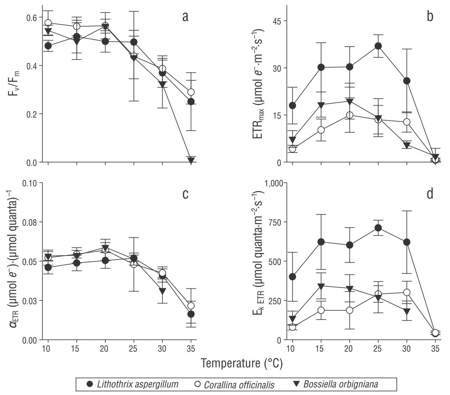
The ETR response to irradiance was similar to that observed in oxygenic photosynthesis (ETR-light curve; Figs. S1, S2); however, ETR values saturated at higher irradiance levels (>300 µmol quanta·m-2·s-1). In general, the highest ETR activity was observed between 20 and 25 °C. Temperature significantly affected Fv/Fm and the parameters derived from the ETR-light curve (P < 0.05; Fig. 2; Tables 1, S1). A stable Fv/Fm of approximately 0.55 from 10 to 25 °C was observed in all species, but it decreased gradually toward higher temperatures to values of ~0.25-0.30 at 35 °C in L. aspergillum and C. officinalis and was close to zero in B. orbigniana (Fig. 2a). Maximum ETR (ETRmax, Fig. 2b) increased from 10 °C to a maximum at 25 °C in L. aspergillum and at 20 °C in the remaining species. Those maxima were shifted by 5 °C toward lower temperatures to the thresholds observed for Pmax. At higher temperatures, ETRmax decreased up to 35 °C in all species. The highest ETRmax values were between 36.9 (±3.6), 14.9 (±5.4), and 19.4 (±5.7) µmol e -·m-2·s-1 in L. aspergillum, C. officinalis, and B. orbigniana, respectively, and decreased 6-fold at 35 °C. The α values, αETR, slightly increased from 10 to 25 °C in all species and then dramatically decreased up to 35 °C, with maximum values between 0.05 and 0.03 µmol e -·µmol quanta·s-1 (Fig. 2c). The Ek ETR values showed mixed responses among species (Fig. 2d). For example, Ek ETR increased from 10 °C to a maximum at 25 °C in L. aspergillum and up to 30 °C in C. officinalis, whereas in B. orbigniana, this maximum was observed at 15 °C. In the optimal temperature range (15-30 °C), the Ek ETR values were ≥600 µmol quanta·m-2·s-1 in L. aspergillum and half of that for the remaining species and were generally higher than those obtained by oxygen evolution.

Figure 2 Maximum photochemical efficiency and photosynthetic parameters describing the electron transport rate (ETR) vs. irradiance curve as a function of temperature in the studied coralline algae. (a) Maximum photochemical efficiency (Fv/Fm), (b) maximum electron transport rate (ETRmax), (c) initial slope of the ETR vs. irradiance curve (αETR), and (d) saturation irradiance for the ETR (Ek ETR). Symbols represent the mean (n = 4) ± SD, except for αETR and Ek ETR for Corallina officinalis at 30 °C (mean ± SD but n = 3). No data for αETR and Ek ETR for Bossiella orbigniana at 35 °C.
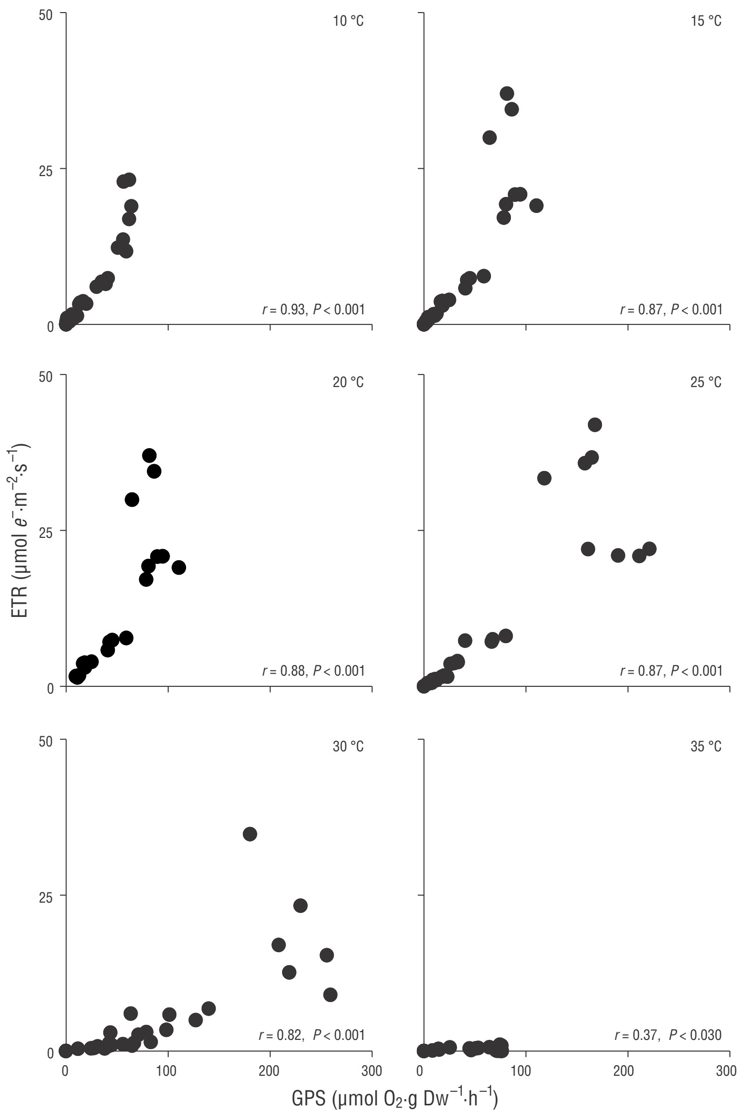
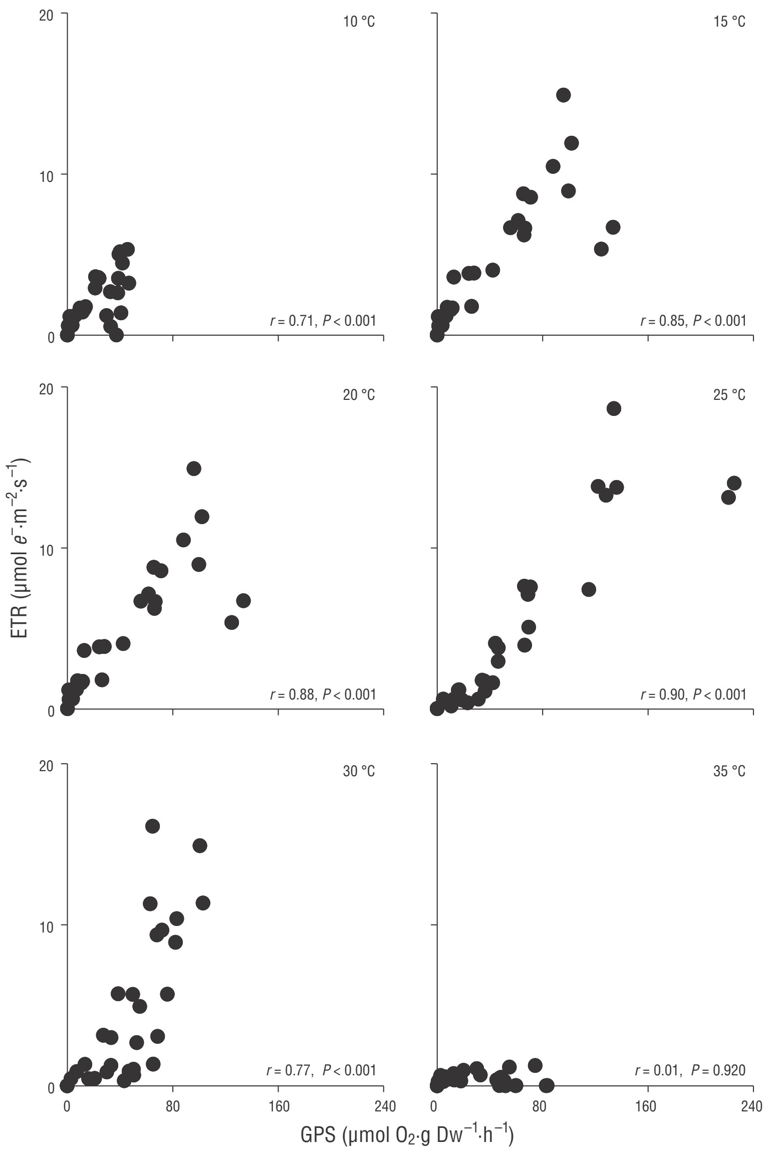
There was a significant linear association (P < 0.05) between GPS and ETR at all temperatures in L. aspergillum (Fig. 3) and from 10 to 30 °C in C. officinalis and B. orbigniana (Figs. 4, 5). In general, correlation coefficients increased with increasing temperatures (up to 0.90) and then decreased at 30 °C. The linear association between GPS and ETR weakened dramatically at 35 °C in L. aspergillum and disappeared in the remaining species. In addition, a deviation from linearity at high irradiances was observed, as ETR generally increased while GPS remained saturated or decreased.

Figure 3 Association between gross photosynthesis (GPS) and the electron transport rate (ETR) in Lithothrix aspergillum as a function of temperature. The P value indicates the significance of the Pearson product-moment analysis (n = 32 per temperature treatment).

Figure 4 Association between gross photosynthesis (GPS) and the electron transport rate (ETR) in Corallina officinalis as a function of temperature. The P value indicates the significance of the Pearson product-moment analysis (n = 32 per temperature treatment, except 30 °C = 24).
DISCUSSION
The results of this study show a high sensitivity of coralline algae metabolism to short-term temperature increases, while this response differed among species. This is in accordance with previous studies on other coralline and fleshy algae (Newell and Pye 1968, Guy-Haim et al. 2016, Vásquez-Elizondo and Enríquez 2016). The observed range of optimal photosynthetic activity and highest P:R ratios correspond to the local annual temperature regime of ~13 °C in winter and ~22 °C in summer (Peña-Manjarrez 2009). The maximum Pmax occurred at higher temperatures (25-30 °C), but P:R ratios and Fv/Fm were already reduced, indicating that optimal growth occurs at lower temperatures. Likewise, these responses may reflect the capacity of the metabolic machinery to rapidly respond to thermal changes not compromising performance.
Photosynthesis and respiration are enzyme-mediated processes that can be limited by substrate availability, and their rates increase with increasing temperatures within the optimal range in a predicted manner (i.e., Q10). This study suggests that Pmax and RD values were strongly regulated by temperature and characterized by differential Q10 (see Newell and Pye 1968). The highest Q10 values, observed in L. aspergillum, may indicate a better adaptation to temperature changes since P:R remained relatively high at 25 and 30 °C, whereas the lowest Q10 (B. orbigniana and C. officinalis) might be attributed to substrate limitation (Eggert 2012) and/or lower thermal tolerance. However, in these 2 species, RD values increase faster than Pmax, indicating an imbalance between carbon loss and gain at elevated temperatures, as well as the production of organic acids and carbohydrates rather than carbohydrates alone (Guy-Haim et al. 2016). The linear increase in RD values suggests a greater thermal tolerance of respiratory enzymatic machinery compared to photosynthetic protein complexes or/and increased respiratory activity as a stress response (Atkin and Tjoelker 2003, Guy-Haim et al. 2016). However, the coralline algal photosynthetic rates were not able to increase in the same manner above 25 °C. In contrast, decreased Pmax at the thermal limits (10 and 35 °C) corresponds to the suboptimal and sublethal portions of the performance curve, typically limited by the amount of enzymes and their activity (Atkin and Tjoelker 2003, Eggert 2012). Other processes, such as photodamage, may have been induced under elevated temperatures (30 and 35 °C), which has been explained as the result of increased light stress, resulting in strong photoinhibition and pigmentation loss (bleaching) (Martin and Gattuso 2009, Martone et al. 2010, Vásquez-Elizondo and Enríquez 2016). Saturation irradiances also decreased at higher temperatures, corroborating that light stress may play an important role in the impairment of photosynthesis during thermal stress. Temperatures of 30-35 °C are above the local thermal regime, but algae might experience these sudden periods of thermal stress on sunny days in the intertidal, which may explain the observed bleaching in other corallines in warmer seasons (Foster et al. 1997, Martone et al. 2010).
The observed species-specific responses are consistent with the photoadaptation of the algae: L. aspergillum does not experience drastic reductions in its carbon balance under thermal changes and has a rather high light adaptation (highest metabolic rates and Ek), while C. officinalis and B. orbigniana show a low light adaptation (lowest metabolic rates and Ek). Coralline algae belong to 2 of 3 main carotenoid profile groups in red algae (lutein and antheraxanthin groups) according to their carotenoid composition, which determines their photoprotection mechanism. The lutein group (L. aspergillum) is characterized by rapid downregulation mechanisms (efficient photoprotection), whereas the antheraxanthin group (B. orbigniana and C. officinalis) is characterized by slow kinetics of Fv/Fm, which is not related to D1 repair (photoinhibition) and is efficient but with slow relaxation (Schubert and García-Mendoza 2008). Such differences may also account for the variance observed in this study. The increased sensitivity of low-light-adapted species in comparison with high-light-adapted species has previously been documented. For example, a linear increase in Pmax and respiration was reported for several moderate and high-light-adapted red algal species, including coralline algae (Cabello-Pasini et al. 2003, Vásquez-Elizondo and Enríquez 2016, Borlongan et al. 2017). In contrast, subtidal algae with low-light adaptation showed optimal activity in a narrower temperature range (Borlongan et al. 2020). Longer exposure times to elevated temperatures and global warming scenarios have yielded diverse responses in coralline algae, but responses also depend on the seasonal acclimation of the algae. For example, Ellisolandia elongata and Lithophyllum incrustans increased photosynthesis with increasing temperature (+3 °C) in winter and summer (Legrand et al. 2018). Similarly, C. officinalis physiology increased during summer in short-term (days) exposure to simulated heatwaves and global change scenarios, whereas longer exposure periods had adverse effects (Rendina et al. 2019). In another study, the photosynthetic activity of high-light-adapted C. officinalis was insensitive to 10 days of temperature changes from 20 to 31 °C, but at higher temperatures, its performance broke down (Guy-Haim et al. 2016). Although the responses observed here may consistently be attributed to physiological adaptations, we cannot rule out that morphological differences (and their implied constraints) among species may have an important impact, which needs to be further explored in future studies.
In this study, a linear association between GPS and ETR was observed under optimal temperatures for photosynthesis. To our knowledge, this is the first study combining simultaneous oxygen evolution and ETR in relation to temperature in coralline algae. However, Sordo et al. (2020), using a single temperature incubation, found a strong association between the GPS and the relative ETR of the rhodolith P. lusitanicum. The responses of the parameters derived from the ETR-irradiance curve are generally well-correlated with the oxygen evolution measurements, but the maximum values of the ETR parameters were shifted by 5 °C toward lower temperatures when compared to the oxygen evolution parameters. Moreover, at 35 °C, the fluorescence signal (∆F/Fmʹ) was almost undetectable, while oxygen evolution still showed activity. In agreement with these results, Hofmann et al. (2012) found a discrepancy between ETR and oxygen evolution parameters for C. officinalis, but only under pH stress. In contrast, a constant relationship between the oxygen and ETR light curve-derived parameters was found for Chondrus crispus, Ulva lactuca, and M. pyrifera (Cabello-Pasini et al. 2000, Cabello-Pasini and Figueroa 2005, Colombo‐Pallotta et al. 2006), but temperature was not tested. Moreover, this linearity between ETR and GPS was observed at non-saturating irradiances (below 200 µmol quanta·m-2·s-1), which also depend on temperature. This is similar to previous results for other macroalgae and seagrasses (see introduction), but exceptions to this also have been reported (Franklin and Badger 2001, Carr and Bjork 2003, Colombo‐Pallotta et al. 2006). Theoretically, the evolution of 1 mol of oxygen releases the flux of 4 electrons needed for carbon fixation, and consequently, a linearity between GPS and ETR is expected to occur (Edwards and Baker 1993). In our approach, we were unable to accurately estimate such a ratio since we used different standardizations for GPS and ETR; therefore, our interpretation is limited to qualitatively assessing both the correlation between GPS and ETR and its sources of variation. The loss of linearity under light saturation (this and other studies) has been explained by the co-occurrence of different electron sinks in excess light to promote photoprotection, such as the Mehler reaction, cyclic electron flow, and photorespiration (Longstaff et al. 2002, Cabello-Pasini and Figueroa 2005). This may be particularly important for red algae lacking a xanthophyll cycle as a main photoprotection mechanism (Schubert and García-Mendoza 2008). An increase in enzymatic activity related to nitrogen assimilation has also been identified as a source of variation within the ETR and GPS relationship (Carr and Bjork 2003, Cabello-Pasini and Figueroa 2005). This might have occurred during periods of optimal performance in this study (20-25 °C), as nutrient uptake increases as a function of temperature.
Under light saturation, high variability in the association between ETR and GPS might also be related to the characteristic presence of phycobiliproteins in red algae and cyanobacteria (Falkowski and Raven 2007). The movement of these complexes along the photosynthetic membranes during and after illumination has strong consequences on the absorption cross section of PSII, which may change the fluorescence signal (Schubert et al. 2011). Indeed, the utility of the classic Fv/Fm approach to estimate photosynthetic electron transport in the coralline Neogoniolithon sp. was found to be unsuitable due to discrepancies between variable fluorescence, non-photochemical quenching, and oxygen evolution, particularly under light stress (Gefen-Treves et al. 2020). In this study, prior to illumination, Fv/Fm had already significantly decreased at 30 and 35 °C, a reduction typically preceded by light stress or a reduction in antenna size, none of which may have occurred with our (dark-adapted) samples. Upon illumination, ∆F/Fmʹ was reduced to half of the maximum photochemical efficiency and less than 0.01 under light saturation, resulting in the ETR being indistinguishable from zero. It is likely that thermal stress activates highly efficient mechanisms of heat dissipation, but more information about fluorescence dynamics under light and thermal stress in coralline algae is crucial to corroborate our observations. Accordingly, the use of ETR under such conditions is discouraged, and other chlorophyll a fluorescence-derived parameters, such as Fv/Fm or Q (PSII pressure), should be preferred for ecophysiological studies (Maxwell and Johnson 2000, Schubert et al. 2011). Our experimental approach uses 2 closely related parameters useful in macroalgal ecophysiology, but caution must be taken when interpreting growth using the GPS and ETR. Even if a close linear relation is found, their increase does not necessarily translate into thallus biomass gain, and in the case of ETR under certain circumstances, into high photosynthetic activity.


 texto en


