1. Introduction
Pathogenic viruses have become a severe threat to health and deaths worldwide. On March 12, 2020, the World Health Organization/WHO (2020) announced the pandemic of coronavirus diseases (Covid-19), which occurred in December 2019. The Covid-19 outbreak is predicted to recover according to the microorganism growth curve. The longer the growth curve of the virus, the longer the outbreak will be. However, without proper regulation and prevention, it would be difficult to predict the length of the growth curve of the virus. This virus can be spread and transmitted through coughing, sneezing, talking, and breathing of an infected person. When mounted on a more miniature carrier aerosol, the combined size is still about 60 nm. However, when mounted on larger carrier aerosols, the combined size can be 100 nm or more significant (Leung & Sun, 2020; Pan et al., 2020).
Many experts recommend reducing the Covid-19 through a healthy, clean lifestyle and using masks. Someone who is required to be in a public place (market, public transportation, and hospital) is always recommended to wear a mask to minimize the transmission of the virus into the respiratory tract (Quan et al., 2017).
Masks are generally made from fiber material. Non-woven materials are particularly suitable for producing synthetic fibers with different morphologies and diameters for air filtration applications. However, synthetic fibers are non- renewable due to environmental issues and biodegradability. The melt-blown process can produce microfibers with 2 μm to 15 μm. They are generally used as a media of non-woven filters. Meanwhile, nanofibers with a diameter around 200 nm - 600 nm produced by electrospinning can be used as filter media for small aerosol filtration, especially for nano-aerosols through diffusion and interception (Leung & Sun, 2020).
The difference in material and the design of antivirus masks will affect the ability of virus and pollutant filtration. The mask's design can include the pores size, the type of material, the number of layers, and the design according to the shape of the face. These parameters are related to filtration efficiency in counteracting certain types of pollutants. The design of masks with the addition of antimicrobial agents or the antimicrobial properties of the polymer itself impacts the inhibition of microbes that infect the respiratory tract. Strict standards are applied to evaluate facemasks/respirators for surgical and healthcare purposes, especially the ability of masks to protect the user from infectious particles. In this case, the antivirus mask prevents droplets and virus particles from infecting people. Therefore, the mask pore size must be produced smaller than the aerosol carrier size and/or smaller than the virus particle size.
In this case, nanometer-sized cellulose fibers (nanocellulose) can be used to produce antivirus masks. Nanocellulose has characteristics that can counteract the spread of the virus through the respiratory tract due to its nanometer size and unique properties. These characteristics include biocompatibility, biodegradable, high mechanical strength, high specific surface area, allows surface functionalization, natural abundance, low cost, and environmentally friendly (Abitbol et al., 2016; Norrrahim et al. 2021b).
Chitin/chitosan nanofiber also has potential as an antiviral mask material for protecting against Covid-19. Chitin/chitosan is produced from the exoskeleton of shrimp and crab through a simple mechanical process after removing protein and minerals. Chitin/chitosan is an abundant and biodegradable biopolymer. The nanofibers obtained had a uniform width of around 10 nm - 20 nm (Ifuku et al., 2010), and the high aspect ratio of chitin/chitosan nanofibers was also known to have antiviral and antibacterial properties (Arkoun et al., 2017).
This review focused on the potency of cellulose and chitin/chitosan-based nanofibers as supporting materials to produce antivirus masks, specifically the Covid-19 virus. Firsthand, cellulose and chitin/chitosan nanofibers and their nanocomposite were described. Secondly, how cellulose and chitin/chitosan nanofibers-based masks could be produced were also explained.
2. SARS-CoV2 dan Covid-19
The coronavirus belongs to the Orthocoronavirinae subfamily, the Coronaviridae family, and the order Nidovirales. This virus is one of the viruses that cause intestinal and respiratory infections in humans. This virus has been considered a pathogen in humans since the Severe Acute Respiratory Syndrome/SARS outbreak in China (Guangdong) in 2002 and 2003. Other coronaviruses also plague the Middle Eastern countries as a cause of the Middle East respiratory syndrome-related coronavirus/MERS- CoV in 2013 (Assiri et al., 2013; Cui et al., 2019). In December 2019, Wuhan in China experienced a new coronavirus outbreak that infected more than 70,000 in the first 50 days of the pandemic. WHO reported that this virus belongs to the β-corona 2B group known as the new coronavirus 2019 (nCov-2019) (Huang et al., 2020; Shereen et al., 2020; Zhu et al., 2020).
The International Committee on Taxonomy of Viruses (ICTV) has called the virus Severe Acute Respiratory Syndrome Coronavirus 2/SARS-CoV2 as the cause of Coronavirus Disease 2019 (Covid19) (Shereen et al., 2020). TEM images show that SARS-CoV2 is generally round in shape with diameters varying from 60 nm - 140 nm (Figure 1). In addition, viral particles have a pointed tip/spike that is typical around 9 nm - 12 nm-the results in the appearance of a virion like a corona/a sun crown.
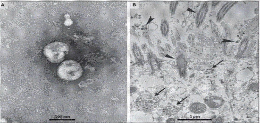
Arrows indicate inclusion bodies formed by viral components
Arrowheads show particles of extracellular virus
Triangles show cilia
Figure 1 Visualization of SARS-CoV2 with TEM, a) extracellular SARS- CoV2 particles and b) SARS-CoV2 in human respiratory tract epithelial cells (Zhu et al., 2020) (Reprinted with permission from Copyright 2020 Massachusetts Medical Society via PubMed Central).
WHO states that the Covid-19 virus can be spread from human to human due to direct contact, also spread of droplets through coughing/sneezing from an infected person. Respiratory infections can be transmitted through various sizes of droplets. Droplet particles with a diameter more than 5 - 10 μm are called respiratory droplets, while droplet particles less than 5 μm in diameter are called nuclei droplets. Therefore, the covid-19 virus is most likely transmitted by respiratory droplets and contact lines (WHO, 2020).
3. Nanofiber filtered mask
A respirator is a facemask designed to give respiratory protection against hostile environments. Respirators can give protection against dust, smoke, pollen, microbes, chemical vapors, and hazardous dust (such as asbestos). There are two respirators, air supplying respirators and air-purifying respirators (APR). APRs have 3 essential components: 1) a facemask, 2) a filter or cartridge filter to remove mists, dust, and smaller particles, and 3) a cartridge filter to remove fumes and chemical gases. Five performance characteristics identified for facemasks are sub-micron particulate filtration, bacterial filtration efficiency, fluid penetration resistance, differential pressure (indicating comfort breathe), and resistance to flammability (Hutten, 2007).
The primary purpose of healthcare and surgical facemasks is to protect the user against colloidal blood splashes and microorganisms when taking care of patients and doing surgery (Hutten, 2007). Simple facemasks cannot prevent infectious diseases, while surgical masks must have at least 80 % bacterial filtering efficiency. Facemasks usually consist of several layers. The layer materials affect the performance of the mask (Akduman & Kumbasar, 2018). When using a coating material with a nanometer structure or increasing the amount of coating material, the filtration efficiency is expected to be higher. On the other hand, the small size of the pores and the increase in the number of layers affect the air permeability, which will impact the supply of oxygen to the respiratory tract. In addition, it is also essential to understand the characteristics of the type of coating material that affect the comfort of its users. The hydrophilic and hydrophobic properties of coating materials also affect the attachment of certain types of pollutants.
Nanofibers can be vital for building filter material in a facemask or respirator. They can increase the filtration efficiency mechanically through the nanostructure (sizeable specific surface area). Therefore, it can effectively increase particles' filtration ability and filter pollutants. Nanofiber filtered mask is also expected to filter PM2.5 particulates (particulate matter with a diameter of 2.5 microns or less) mechanically and filter and/or kill bacteria/viruses through nanocomposite structures with an antimicrobial agent. Nanofiber filtered masks consist of layers with lightweight and flexible structure and morphology, thus providing comfort for its users.
The different processes for preparing non-woven filters (such as facemask) will produce different filters even from the same material. The primary process for preparing a non- woven filter can be divided into dry and wet processes. In the dry process, the webs are formed in an air medium. In comparison, the webs are formed in water in the wet process. There are five dry processes: dry-laid (carding operations), air- laid, spun-bonded, electrospun, and melt-blown. Meanwhile, the wet-laid process is similar to the papermaking process (Hutten, 2007).
Nanofibers filtered masks for protecting against Covid-19 must meet the minimum requirements for filtration by the Food and Drug Administration (FDA) under ASTM F2100-11. According to ASTM F2100-11, the performance of medical facemask material is based on testing particulate filtration efficiency, resistance to penetration by synthetic blood, differential pressure, flammability, and bacterial filtration efficiency. In addition, medical filtration mask material must be designed based on the properties of barrier performance of the medical mask material, including Level 1 Barrier, Level 2 Barrier, and Level 3 Barrier. Meanwhile, Table 1 shows medical filtration mask material requirements based on ASTM F2100-11. The N95 respirator has a filter efficiency based on the particles being filtered. Therefore, every biological particle is expected to be filtered with an efficiency not less than that of aerosol testing (at least 95 % efficient for N95 filters) (Akduman & Kumbasar, 2018).
Table 1 Medical filtration mask material requirements based on ASTM F2100-11.
| Characteristic | Level 1 Barrier | Level 2 Barrier | Level 3 Barrier |
|---|---|---|---|
| Bacterial filtration efficiency (%) | ≥95 | ≥98 | ≥98 |
| Differential pressure (mm H2O/cm2) | <4.0 | <5.0 | <5.0 |
| Sub-micron particulate filtration efficiency at 0.1 microns (%) | ≥95 | ≥95 | ≥95 |
| Resistance to penetration by synthetic blood | 80 | 120 | 160 |
| Flame spread | Class 1 | Class 1 | Class 1 |
Furthermore, WO 2016/128844 A1 patent (Majmi et al., 2016) described the process of producing nanofiber facemasks. Facemasks were made with three layers, one nanostructured layer in the middle and two non-woven covering layers. The nanofiber layer was made using needleless electrospinning. Polymer solutions used for nanofibers included polyolefins, polyamides, polyesters, ether and cellulose esters, cellulose acetate, polyvinylidene fluoride, polyaerylonitrile, polyvinyl alcohol, polyether sulfon, nylon, polystyrene, polyaerylonitrile, polycarbonate, chitosan, mixtures. Meanwhile, two cover layers were polypropylene, polyester, polyethylene, polyamide, polyurethane, or mixed polymers.
Nanofiber membrane has a low basis weight, small pore size, and high permeability. Moreover, nanofiber cellulose- based membranes are unique with good interconnectivity of pores, high specific surface area (in the range from 1 m2/g to 100 m2/g depending on intra-fiber porosity and fiber diameter), and the potential to combine active chemistry or functions at the nanoscale (Akduman & Kumbasar, 2018). Therefore, cellulose nanofibers are not independently used purely as anti-Covid-19 masks but can also be added with the active agent (antivirals) or other composites to improve their performance and mechanical properties.
Filter technical requirements aim to balance the three primary parameters of filter performance, namely pressure drop, filter life, and filter efficiency. In this context, cellulose nanofibers can increase the efficiency of particle capture and increase the pressure drop associated with the thickness of the nanofiber on a primary filter substrate (Wang et al., 2015).
The high surface area to volume ratio of nanofibers is likely to increase particle deposition on the fiber surface. Therefore, it impacts improving filter efficiency with a relatively low- pressure drop. The high filtering efficiency effectively prevents ambient PM2.5, which entering the human body. The mechanism for making nanofiber masks can be seen in Figure 2.
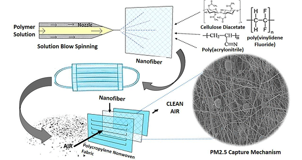
Figure 2 Mechanisms for making nanofiber masks (Tan et al., 2019) (Reprinted with permission from Tan et al., 2019. Copyright 2020 American Chemical Society).
4. Cellulose nanofibers
Cellulose is the main component for building plant cell walls (about 35 % - 50 % of the dry weight of plants). In nature, cellulose is associated with other polysaccharides such as hemicellulose and lignin (Fahma et al., 2010). Cellulose is a linear homopolysaccharide consisting of β-D-glucopyranose units connected by β-1-4 bonds (Brännvall, 2007).
Cellulose nanofibers, also known as nanocellulose, are cellulose-based nanostructures whose diameters are on the nanometer scale (1 nm - 100 nm). Nanocellulose can be isolated from various lignocellulosic plants and biomass with chemical treatments using acid hydrolysis and mechanical treatments using a combination of ultrafine grinding and ultrasonication (Fahma et al., 2010; 2011; 2016; 2017a; 2019; Lisdayana et al., 2018; Wahyuningsih et al., 2016). In addition, nanocellulose can be produced using the electrospinning method of cellulose solution (Kim et al., 2006). Bacterial cellulose can also be categorized as nanocellulose because of the nanostructured gel produced (Klemm et al., 2009).
Bacterial cellulose is a cellulose fiber produced by Acetobacter xylinum, Agrobacterium, Aerobacter, Achromobacter, Azotobacter, Rhizobium, Sarcina, Alcaligenes, Pseudomonas, Dickeya, Rhodobacter, and Salmonella (Lustri et al., 2015). Acetobacter xylinum (optimal at room temperature, pH 3.5 - 4) is considered the most efficient producer of bacterial cellulose. Bacterial cellulose produces very pure cellulose in nanofibers with unique physical properties (Sani & Dahman, 2010). The nanostructure of bacterial cellulose can provide exceptional mechanical properties. The tensile strength of bacterial cellulose is around 2 MPa in wet conditions and 200 MPa - 300 MPa in dry conditions (Gatenhol & Klemm, 2010). In addition, bacterial nanocellulose is widely used in biomedical science because of its high biocompatibility and purity. According to Yao et al. (2017), bacterial nanocellulose has very crystalline properties with high intrinsic mechanical properties. Figure 3 shows cellulose nanofibers from oil palm empty fruit bunches (OPEFBs) and bacterial cellulose.
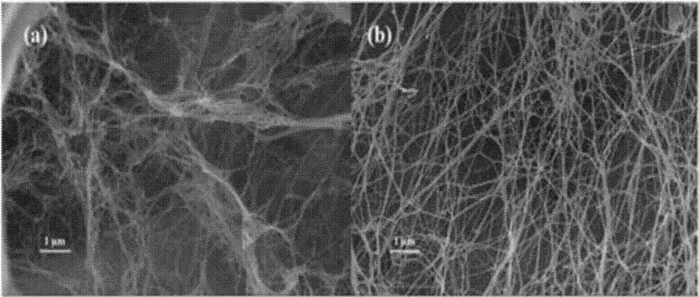
Figure 3 SEM images of nanocellulose from, a) OPEFBs and b) bacterial cellulose (Fahma et al., 2019) (Reprinted with permission from Copyright 2020 Taylor & Francis).
Nanocellulose is wholly dispersed in the polar polymer matrix, such as polyvinyl alcohol (Fahma et al., 2017b). Meanwhile, nanocellulose is not easily dispersed homogeneously in a non-polar polymer matrix because of its hydrophilic nature. In addition, cellulose hygroscopicity causes dimensional instability of the composite shape. The hydrophilic nature of cellulose is caused by the large number of hydroxyl groups located on the surface of cellulose fibers. So, it is necessary to chemically modify the surface of nanocellulose using various reagents, for example, the acetylation process, which produces cellulose acetate (CA). Acetylation replaces hydroxyl groups in cellulose with less hydrophilic acetyl groups (Fahma et al., 2014a; 2014b).
4.1. Cellulose nanofiber mask for virus filtration
Nanocellulose is a promising material for the development of nanofiltration masks. The high specific surface area allows for the adsorption of various microbial species. Its inherent porosity can also separate various molecules and maintain microbial objects. In addition, the presence of large OH groups in nanocellulose provides a unique surface modification to increase its filtration efficiency through the formation of affinity interactions for microbes (Norrahim et al., 2021a).
Size exclusion is a concept used to screen microbes depending on their size or "hydrodynamic volume" in solution. When different molecules are combined in the matrix, small molecules diffuse into the pores and slow their flow through the column. Meanwhile, large molecules (or having an enormous hydrodynamic volume) do not penetrate the pores and are eluted in the empty volume of the column. Therefore, the acidity (pH) and ionic strength of the load buffer will significantly impact the retention of various specimens. In addition, electrostatic interactions with ions in solution causes charged pollutants to have a double layer of electrical charge on their surface. The principle of affinity using adsorption is also used to remove pollutants based on electrostatic interactions between membrane functional groups and pollutants. Therefore, this principle can be used as a basis for counteracting charged particles (Norrahim et al., 2021a).
Nanocellulose has been known as a fiber capable of separating viruses (Table 2). The nanocellulose filter succeeded in trapping Swine Influenza Virus (SIV) particles measuring 80-120 nm, similar to SARS-CoV2 (Metreveli et al., 2014). Nanocellulose needs to be surface modified to become cationic to increase the efficiency of virus screening. Corona and several other viruses have a negatively charged effectively trapped due to electrostatic attraction between negatively charged viruses and positively charged nanocellulose (Norrrahim et al., 2021b).
Table 2 Nanocellulose as a virus filter material.
| Material | Result | Ref. |
|---|---|---|
| Nanocellulose filter paper | Non-woven filter paper with a thickness of µm containing nanocellulose fibers can remove virus particles based on the Size-Exclusion principle, a log10 reduction value (LRV) 6.3 | Metreveli et al. (2014) |
| Citric acid crosslinked nanocellulose | Paper filters can remove tracer particles as small as 20 nm, including viruses. | Quellmalz and Mihranyan, (2015) |
| Nanocellulose-based mille-feuille filter paper | Although their flux was generally lower, the 33 µm filter exhibited more robust virus removal properties and better throughput properties than the 11 µm filter. | Manukyan et al. (2011) |
| Nanocellulose based filter paper | Nanocellulose filter paper helps remove endogenous rodent retroviruses and retrovirus-like particles during the production of recombinant proteins. This study tested a xenotropic murine leukemia virus model. | Asper et al. (2015) |
| Nanocellulose-based nanoporous filter paper | Filters showed 5-6 logs of virus clearance of ΦX174 (28 nm) or MS2 (27 nm) phages. The mille-feuille filter paper manufacturing offers the possibility of producing cost-efficient virus removal filters with performance capabilities for processing plasma-derived immunoglobulins and recombinant monoclonal antibodies. | Wu et al. (2019) |
| Nanocellulose-based size-exclusion filter | The nanocellulose-based filters exhibit high virus retention capacities (> 4 log10) and high flow rates (approximately 180 Lm-2h-1), so they have the potential to be developed as cost-effective virus removal filters for upstream bioprocesses. | Manukyan et al. (2019) |
| Nanocellulose-based filter paper | Sequential filtration through an 11 µm prefilter and 33 µm virus removal filter paper allowed high virus removal capacity. | Manukyan et al. (2020) |
| Nanocellulose-based filter paper | Nanofiltration with 11 µm thickness (prefilter) and 22 µm (virus filter) was able to remove viruses from plasma-derived human serum albumin (HAS) in bioprocessing as an alternative to virus inactivation method. | Wu et al. (2020) |
Three charging techniques are used to make filter media with electrostatic methods: electrostatic fiber spinning, corona charging, and tribocharging. Each technique is used for different types of polymers. Corona charging is suitable for charging mono- polymer fibers or fiber fabrics or blends. Tribocharging is only suitable for charging fibers with different electronegativity. Meanwhile, electrostatic fiber spinning combines polymer charging and fiber spinning as a one-step process. Two different fibers after tribocharging have a higher filtration efficiency than corona-loaded polypropylene fibers (Tsai et al., 2002).
Electrospinning is one of the most common methods to produce nanofibers which is relatively easy. Electrospinning produces nanofibers with unique characteristics, such as high surface and volume ratio, low basis weight, high permeability, relatively uniform fiber size, and small pore size. Therefore, this method is appropriate for nanofiltration applications with tiny particles (Leung & Sun, 2020). This method has been successfully used to produce nanofibers based on polyvinyl alcohol (PVA), polyurethanes, polypyrrolpolyacrylonitrile-co- vinyl acetate, polyacrylonitrile (PAN) and dimethylformamide, alumina, polypropylene, polyamide-6, and activated carbon, as well as several nanocomposite polymers (Ahne et al., 2019).
Electrospinning has also been used by Omollo et al. (2016) to produce high-efficiency filtration using nanofiber cellulose acetate. Cellulose acetate (CA) is dissolved in trifluoroacetic acid and electrospun into non-woven polypropylene. In order to form a three-layered polypropylene/cellulose acetate/polypropylene filter, an upstream layer of polypropylene non-woven material was added (Figure 4). The results showed that increasing the thickness of the CA nanofiber layer could improve filtering efficiency. Furthermore, filter performance showed that PP/CA/PP material could be used as high-efficiency filtration material. The CA nanofibers added to PP non-woven materials to form PP/CA/PP-6 filter have diameters ranging from 300 nm to 400 nm, more significant than electrospun (120 nm - 130 nm).
Several supporting materials can be used to improve the performance of cellulose acetate fibers to produce multilayer filters. Kim et al. (2015) researched CA/MMT nanofiber composites using electrospinning. The results, the average diameter of composite nanofiber with 18 % CA/MMT solution in acetic acid/water (75/25, w/w) solvent ranged from 150 nm to 350 nm. The diameter of the obtained composite- nanofibers decreased (around 90 nm) with increasing MMT (Figure 5).
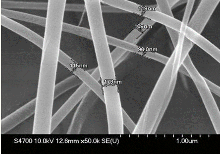
Figure 5 SEM image of CA/MMT composite nanofibers with 9 wt% MMT (Kim et al., 2015) (Reprinted with permission from Copyright 2020 Hindawi).
Su et al. (2010) examined the nanofiltration membrane of cellulose acetate. The results show that the membrane pores produced are microscopic. The existence of heat treatment (60 minutes at 60° C and 20 minutes at 95° C) can reduce the average size of the radius of the membrane pores from 0.63 nm to 0.30 nm. Ahne et al. (2019) have studied the key parameters that affect the filtration quality factors in nanocellulose acetate filters, including solvent concentration, voltage, tip distance to the collector, and deposition time during electrospinning. The cellulose acetate concentration tested was 10 wt % - 20 wt % using N, N-dimethylacetamide (DMAc), and acetone. This study used the ratio of cellulose acetate: solvent 2:1, a constant feed flow rate of 0.6 ml/h, electrospinning voltage around 8 kV - 12 kV with a distance of 10 cm -15 cm, and a deposition time of 30 minutes. The average fiber diameter ranges from 175 nm - 890 nm, and CA concentrations below 15 % caused beads formation.
Su et al. (2010) examined the nanofiltration membrane of cellulose acetate. The results show that the membrane pores produced are microscopic. The existence of heat treatment (60 minutes at 60° C and 20 minutes at 95° C) can reduce the average size of the radius of the membrane pores from 0.63 nm to 0.30 nm. Ahne et al. (2019) have studied the key parameters that affect the filtration quality factors in nanocellulose acetate filters, including solvent concentration, voltage, tip distance to the collector, and deposition time during electrospinning. The cellulose acetate concentration tested was 10 wt % - 20 wt % using N, N-dimethylacetamide (DMAc), and acetone. This study used the ratio of cellulose acetate: solvent 2:1, a constant feed flow rate of 0.6 ml/h, electrospinning voltage around 8 kV - 12 kV with a distance of 10 cm -15 cm, and a deposition time of 30 minutes. The average fiber diameter ranges from 175 nm - 890 nm, and CA concentrations below 15 % caused beads formation.
Akduman (2021) combined electrospun nanocellulose acetate (CA) and polyvinylidene fluoride (PVDF) coated with polypropylene spun to produce a facemask layer that meets the N95 respirator requirements. The nanofiber layer produced showed higher mechanical filtration efficiency compared to conventional facemasks. In addition, the effects of electrospun CA and PVDF nanofiber's diameter on the thickness and pore size of the mats were compared in terms of filtration performance. The diameter of the fibers decreased with decreasing concentrations of PVDF polymers (236.50 nm to 142.59 nm) and CA (319.02 nm to 264.02 nm).
Some studies have been carried out to observe the performance of nanocellulose acetate as a nanometer-sized particle filter in the air. Chattopadhyay et al. (2016) examined electrospun cellulose acetate for aerosol filters (solid and liquid) by examining the effect of fiber diameter, thickness, and solidity of the nanofiber membrane electrospinning method. Electrospinning used varies with fiber diameter (0.1 µm - 1 µm), filter thickness (7 µm - 51 µm), and solidity (0.1 - 0.2). The results showed that the diameter of the obtained fibers ranged around 0.1 μm - 24 μm with filter thickness ranging around 7 μm - 51 μm. This study also showed that the electrospun nanofiber filtration performance was slightly better than commercial glass fiber filters. Solid aerosol particles tend to penetrate the first few layers of filter media depending on the fiber diameter.
Li and Gong (2015) prepared nanofiber masks by dissolving polysulfone (18 % concentration) in DMAc/acetone (9:1) with intense agitation. The solution was stored for 24 hours without agitation at room temperature to remove air bubbles. The 18 % cellulose solution was inserted into the syringe with a metal needle connected to a high voltage power supply, about 13 KV with a needle and aluminum foil distance (13 cm). The polymer solution was fed using a syringe pump at a constant rate of 0.4 mL/h. Nanofibers were collected on the surface of PP non-woven as a collector. The results indicated that the nanofiber mask could efficiently filter out the PM2.5 particles and simultaneously preserve excellent breathability. Figure 6 shows the nanofibers on the non-woven fabric.
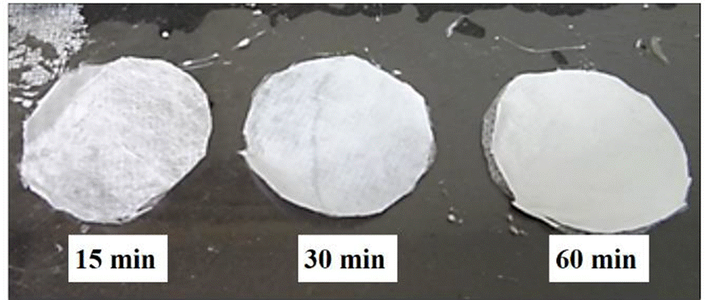
Figure 6 Nanofibers on the non-woven fabric (Li & Gong, 2015) (Reprinted with Li & Gong, 2015. Copyright 2020 Hindawi).
4.2. Antimicrobial cellulose nanocomposite mask for Covid-19
The quality of protective masks depends on the dryness and hydrophobic nature of the protective layer (outer layer). If this layer is not sensitive to microorganisms, it can cause health risks and the easy growth of pathogenic viruses/bacteria under humid conditions. Therefore, nanofiber masks need to be composited with antibacterial/antiviral agents to minimize bacterial infections or SARS-CoV-2 in the respiratory tract. Therefore, the use of antimicrobial masks is considered an essential thing to consider for preventing Covid-19.
Hiragond et al. (2018) submitted that antimicrobial masks had become a concern in the development of medical textile fabrics. Silver (Ag) is an antimicrobial material that has been widely used in many household products. The Ag nanoparticles (nanosilver) size is significantly beneficial for more effective interactions with bacterial/viral membranes. Nanosilver, in this case, can easily cross the lipid bilayer and plasma membrane and enter the cytoplasm. The entry of Ag nanoparticles into the cytoplasm will eventually destroy bacterial cells through attacks on DNA. In fighting the SARS- CoV-2 virus, AgNPs inhibited the glycine and alanine of the S- protein and other proteins. S-protein is the most critical component because it causes rapid replication in the host body (Ahmed et al., 2021).
Chunju et al. (2003) developed a cellulose fiber method for antivirus and antibacterial masks. They examined the solvent method for preparing cellulose and containing antivirus. Chunyan (2009) examined the manufacturing of filtration masks from bacterial cellulose. This mask consists of three layers, namely two layers of bacterial cellulose diaphragm and one bacterial cellulose stenlizing layer. Bacterial cellulose stenlizing layer must be infiltrated on nanosilver solution after freeze-drying. Filters made from bacterial cellulose were compiled with nanosilver measuring around 20 nm. Nanosilver materials used are silver nitrate, silver chlorate, silver citrate, or silver oxalate. Bacterial cellulose was washed with a 0.5 % alkaline solution and stored at 2° C - 4° C. Nanosilver dissolved in water to form nanosilver concentrations (0.001 mol/L ∼ 0.050 mol/L) to be infiltrated in bacterial cellulose. The infiltration process was stopped at the desired concentration reached. Next, freeze-drying was done to obtain the stenlizing layer. This technology is more superficial, with low production costs, high adsorption capacity, high antibacterial and antiviral activity than Ag can effectively reduce resource consumption and preserve the ecological environment.
Hamouda et al. (2021) added AgNPs in cotton and polyester knitted fabrics blended with spandex yarns for mask application. The count and ratio of different spandex yarns will reduce the porosity and air permeability of the fabric. The combination of a three-hybrid layers' mask made of polyester fabric on the outer layer with 100% cotton fabric in the inner layer showed high air permeability and breathability. In addition, wearing a mask during activities did not significantly affect the wearer's blood oxygen saturation and heart rate. In this regard, AgNPs have low cytotoxicity and considerable efficiency in inhibiting SARS-CoV-2. However, repeated inhalation of nanosilver poses risks (Christensen et al., 2010). Many studies have reported that nanosilver is toxic to bacteria (McShan et al., 2014; Naidu et al., 2015; Pulit-Prociak et al., 2015). Therefore, it is necessary to have toxicity data on absorption (particles or ions), information on gene toxicity, and further information on toxicity after inhalation exposure to agglomeration size and status as unaccounted in the workplace (Christensen et al. 2010). In producing bacterial nanocellulose composite masks with nanosilver, it is necessary to consider the level of toxicity that can endanger health.
Copper oxide (CuO) can also be compiled with a breathing mask prepared from nanofibers to add antimicrobial properties (Hashmi et al., 2019). CuO has a more economical production level than nanosilver, gold nanoparticles, and copper nanoparticles. Copper (II) oxide is hydrophilic and stable. The results showed that PAN (Polyacrylonitrile)/CuO nanofibers were successfully electrospinning with various concentrations of CuO in PAN nanofibers. The addition of CuO nanoparticles was able to add strength to PAN nanofibers. Significantly, the tensile strength of PAN nanofibers with an additional 1 % CuO nanoparticles could increase up to 8.43 MPa. Morphological properties showed uniformity and smoothness of nanofibers production (beads-free nanofibers). The produced nanofibers had excellent antimicrobial activity. The similarity characteristics of PAN and hydrophilic nanocellulose can be compiled with antimicrobial agents for antiviral properties. Meanwhile, the toxicity test is used to determine the toxic effect of the material when inhaled directly by humans. As a result, pure PAN nanofibers have good biocompatibility and lower toxicity. On the other hand, the increased addition of CuO leads to a toxic effect which is recommended <1% based on tests on cells (NIH3T3) (Hashmi et al., 2019).
5. Chitin/chitosan nanofiber mask for Covid-19
5.1. Chitin/chitosan nanofiber
Chitin is a high molecular weight polymer with a β (1,4)-N- acetyl glycosaminoglycan-repeating structure, while chitosan is a deacetylated chitin. Chitin is very crystalline with a strong hydrogen bonding with high binding energy. These properties are due to its linear structure with two acetamide and hydroxyl groups arranged in an antiparallel fashion as nano-sized chitin nanofibers. The nanofibers diameter is around 2 - 5 nm with a length of about 300 nm embedded in a protein matrix. Chitin is found in crab shells, shrimp, lobster, insects, sea diatoms, algae, mushrooms, and others (Zhang & Rolandi, 2017). Crab and shrimp shells have a hierarchical structure consisting of nanofibers (Figure 7). Chitin consists of very uniform nanofibers with a diameter of 10 nm - 20 nm, uniform structure, and high aspect ratio (Ifuku & Saimoto, 2012).
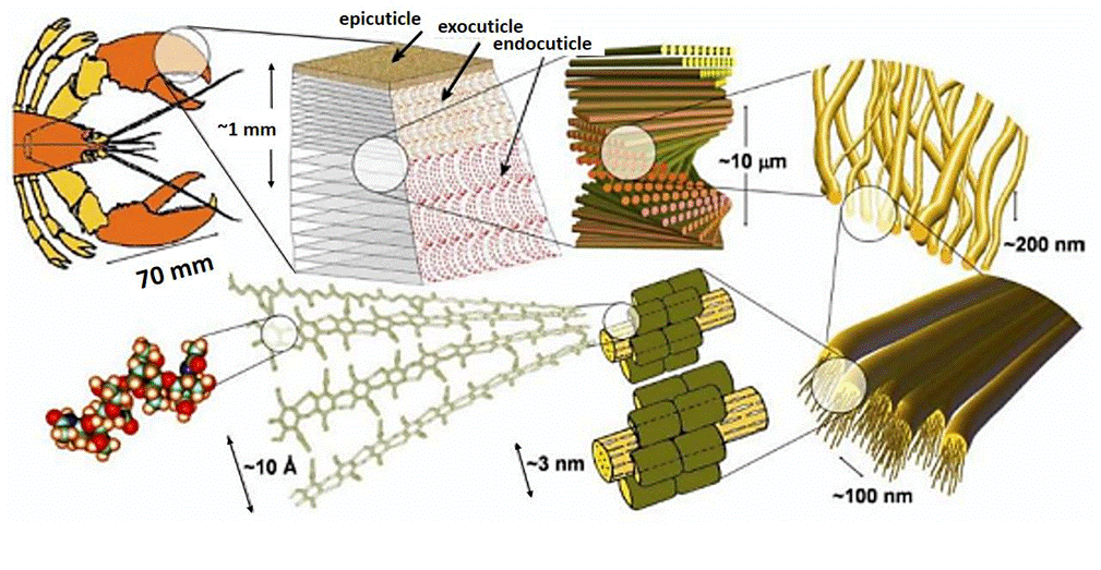
Figure 7 Schematic presentation of nanostructure of crab shells (Zhang & Rolandi, 2017) (Reprinted with permission from Zhang and Rolandi, 2017. Copyright 2020 Royal Society of Chemistry).
According to Ifuku (2014), chitin nanofiber has a diameter of up to 10 nm (Figure 8). Chitin powder and commercial chitosan can be easily converted into nanofibers by mechanical treatment through grinding and high-pressure waterjet systems; this is caused by chitin powder and chitosan consisting of nanofiber aggregates. Acidic conditions are a crucial factor in mechanical fibrillation. Increasing the surface property of chitin nanofiber can be modified by graft polymerization, methylation, chlorination, naphthaloylation, phthaloylation, acetylation, deacetylation, and TEMPO- mediated oxidation. In addition, partially deacylated chitin nanofiber has a highly reactive amino group. This group is vital for determining the role of chitin and cellulose.
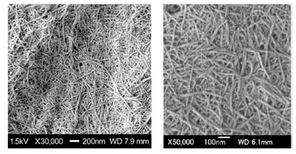
Figure 8 SEM images of chitin nanofibers from crab shells after grinding (Ifuku, 2014) (Reprinted with permission from Ifuku, 2014. Copyright 2020 MDPI).
5.2 Chitin/Chitosan nanofiber mask
The manufacture of chitin/chitosan nanofiber masks for Covid-19 is almost the same as air filtration. Therefore, chitosan as a nano mask media is advantageous as an air filter. Desai et al. (2009) made nanofibers-based filter media from chitosan/PEO electrically to a non-woven polypropylene substrate. The efficiency of nanofibers filtering was closely related to the size of the fiber and the content of chitosan. Liu et al. (2019) developed a transparent air nanofilter from chitosan nanofibers (Figure 9). These bilayer air filters consisted of poly (methylmethacrylate) (PMMA)/polydimethylsiloxane (PDMS) superhydrophobic fibers as a barrier to moisture intake. Chitosan super hydrophilic fiber was used for the efficiency of capturing PM (particulate matter) of more than 96% optical transmittance of 86 %. In addition, this filter was able to capture PM2.5 at a higher rate (more than 98.23 %). The nanofilter membrane also had antibacterial properties against Escherichia coli (96.5 %) and Staphylococcus aureus (95.2 %). The production process of air filters is shown in Figure 10, and the transition behavior of airflow under high humidity in Figure 11.

Figure 9 Different transparencies of chitosan-PDMS/PMMA nanocomposite-based air filters at different optical transmittance (16 %, 37 %, 54 %, 71 %, and 86 %) (Liu et al., 2019) (Reprinted with permission from Liu et al., 2019. Copyright 2020 Elsevier).
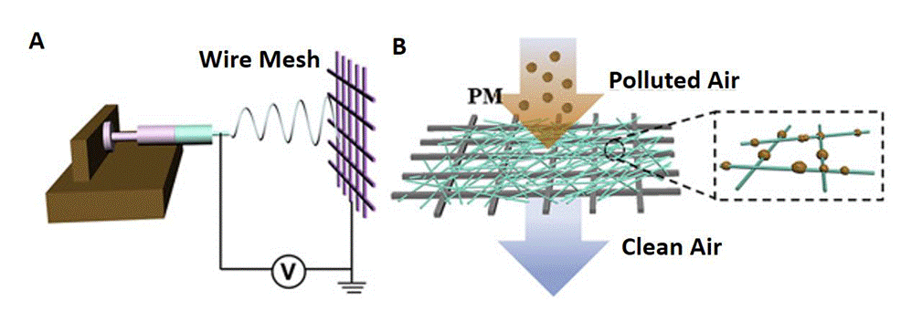
Figure 10 The production process of air filters, a) Schematic of air filter production using Electrospinning, and b) Illustration of the filtration process of the fibrous membrane (Liu et al., 2019) (Reprinted with permission from Liu et al., 2019. Copyright 2020 Elsevier).
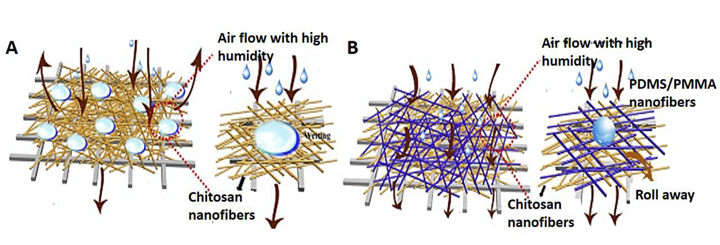
Figure 11 Transition behavior of airflow pass. A: through pure chitosan nanofiber-based filters. B: through chitosan-PDMS/PMMA nanocomposite-based filters under humidity conditions (Liu et al., 2019) (Reprinted with permission from Liu et al., 2019. Copyright 2020 Elsevier).
Shahvaziyan et al. (2013) utilized chitosan nanofibers as an air pollutant filtration material by electrospinning. The optimal conditions for making filters were obtained with a mass density of 1.7, a feeding rate of 1 ml per minute, a voltage of 15 kV, and a distance of 13 cm between the opening and the collecting plate. Based on tests on pollutants, commercial masks could not remove almost all pollutant metals, but masks made from chitosan nanofiber could filter a large number of pollutant metals such as Pb, Cd, Sb, etc.
Mohraz et al. (2019) examined the effect of the uniformity and diameter of electrospun polyurethane/chitosan nanofibers as filtration and antimicrobial activity against nanoaerosol bacteria. The average nanofibers diameter decreased significantly with increasing chitosan content in the polyurethane/chitosan mixture and voltage. The polymer flow rate, the distance of the needle tip to the collector, and the diameter of the average nanofibers show a significant positive correlation. An antibacterial test using the disk diffusion method showed that the polyurethane nanofiber/chitosan electrospun had good antibacterial activity against Escherichia coli.
5.3 Antiviral chitin/chitosan nanofiber mask against Covid-19
Nguyen et al. (2014) investigated the antimicrobial properties of Ag nanoparticles (AgNPs) nanocomposite sheets with chitin-nanofibers (CNFS). The results showed that Ag NPs immobilized in CNFS gave a much higher antimicrobial activity against Escherichia coli, Pseudomonas aeruginosa, and human influenza A viruses (A/PR/8/34 (H1N1). Furthermore, high concentrations of AgNPs immobilized in CNFS could significantly reduce the virus in logs of 10 CFU/ml, while CNFS provided a minor reduction in influenza A viruses. In addition, Sun et al. (2012) investigated the use of chitosan nanoparticles loaded with nucleocapsid proteins from bovine coronavinis (BCV N). This process used the ionic crosslinking method with sodium tripolyphosphate as a crosslinking agent. Antiviral activity of chitosan nanofiber and HTCC or (N - [(2- hydroxy-3-trimethylammonium) chitosan chloride) was investigated by Bai (2012). Electrospun combination of HTCC/graphene in water can eliminate Porcine Parvovirus (PPV) up to 99 %. Meanwhile, chitosan nanofibers were only able to eliminate 50 % of viruses. The average diameter of electrospun chitosan fiber ranges from 80 nm - 130 nm. The smaller diameter of the fiber increased the surface area to increase the binding of PPV. The diameter of the HTCC/graphene fiber increased with increasing concentration.
Furthermore, the hydrophobicity of graphene and the high HTCC content could bind 95 % PPV. Therefore, HTCC/graphene nanofibers can be used as nano-membranes/microfiltration to remove viruses by adsorption (Bai et al., 2013). In addition, the addition of affinity ligands to electrospun fibers was also able to increase the binding of viruses such as peptide ligands to eliminate PPV (Heldt et al., 2008). Adding peptides (WRW and KYY) to the chitosan amine group was thought to increase the binding ability of chitosan nanofibers compared to HTCC nanofibers. WRW and KYY peptides are positively charged with two hydrophobic amino acid groups (Bai, 2012).
In addition, nano/microspheres of N-(2-hydroxypropyl)-3-trimethyl chitosan (HTCC-NS/MS) were studied as adsorbents of human coronavirus NL63 (HCoV-NL63), mouse hepatitis virus (MHV), and HCoV-OC43 particles (Ciejka et al., 2017).
Cationically-modified genipin crosslinked chitosan nano/microspheres enabled the reversible adsorption of human coronavirus particles NL63 (HCoV-NL63) without losing their infectivity. Furthermore, the adsorption capacity of HTCC- NS/MS correlated well with the antiviral activity of soluble HTCC against certain viruses. Importantly, HCoV-NL63 particles could be desorbed from the surface of HTCC- NS/MS with a high ionic strength salt solution with retention of virus virulence. Furthermore, increased cationization of polymer particles can increase the adsorption of HCoV-NL63. The suggests that ionic strength- induced viral desorption suggests that the virus-polymer interaction is due to electrostatic Coulomb attraction. Therefore, HTCC-NS/MS has excellent potential in detecting, removing, separating, separating, and/or purifying the HCoV-NL63 coronavirus.
6. Future directions
The nanotechnology approach is expected to innovate biopolymer-based nanofiltration masks against the coronavirus. Nanocellulose and chitin/chitosan are promising biopolymers to be developed into environmentally friendly nanofiltration masks. The utilization of the biopolymer is based on its ability to filter or separate virus particles. In addition, the addition of metal nanoparticles in the mask design is expected to increase the filtration efficiency. Some things need to be considered when designing an antiviral mask based on nanocellulose, chitin/chitosan, or the addition of active ingredients, including:
Modifying nanocellulose to be positively charged is thought to increase the efficiency of nanofiltration in separating negatively charged coronaviruses.
Cationically modified chitosan is also possible to separate coronavirus particles.
The addition of active ingredients (e.g., nanosilver) can increase the effectiveness of nanofiltration masks against the coronavirus.
The combination of nanocellulose, chitin/chitosan, and active ingredients is a strategy to improve the performance of nanofiltration masks. Therefore, it is necessary to research the percentage of each ingredient added to the mask design.
In designing masks, it is crucial to pay attention to the level of toxicity of each constituent and the use of chemicals.
The pores size of the mask must be designed to be smaller than the size of the coronavirus. On the other hand, the minimal size of the pores will hinder the supply of oxygen to the respiratory tract.
7. Conclusions
Nanofiber-based masks can be used as filtration masks to prevent the Covid-19 virus. Nanofiber masks can be made with fiber sizes or pores more minor than the Covid-19 virus diameter to below 80 nm. Potential nanofiber materials for the Covid-19 nano mask include bacterial nanocellulose and/or acetate, cellulose nanofiber, and chitin nanofiber. This mask can offer several advantages, including biodegradable, biocompatible, good mechanical properties, high filtration ability (depending on the nanometer pores and fibers), large abundance, and inexpensive. In addition, nanofiber masks can be composited with nanometer-sized active materials (such as CuO, nanosilver, nano gold, natural materials, etc.) to improve the performance of nanofiber masks in killing bacteria or pathogenic viruses (specifically Covid-19 virus). The selection of active ingredients of the composite must consider the toxicity.


 nueva página del texto (beta)


