Introduction
Due to the high cost represented by dissection, grinding and Chemical analysis, alternative methods for determining the composition of the carcass and body of farm animals have been studied (Silva et al. 2015, Silva et al. 2016, Silva 2016). Among these, indirect methods are preferred because they are considered non-invasive (Scholz et al. 2015), are easier and cheaper to implement and can be applied to live animals (Silva et al. 2015, Silva 2016). Among current techniques, the most notable are ultrasound (Aguilar-Hernández et al. 2016), biometric measures (Fonseca et al. 2017, Bautista-Diaz et al. 2017) and digital image analysis (Gomes et al. 2016).
Some authors have reported that the use of ultrasonography is a non-invasive technique that can predict the amount of muscle, bone and fat in meat and wool sheep breeds (Silva et al. 2006, Ripoll et al. 2009) and recently in hair ewes (Aguilar-Hernandez et al. 2016). Moreover, ultrasound measurements (USM) may contribute to predicting subcutaneous body fat, and the characteristics of carcass tissues such as the Longissimus dorsi area and depth in live farm animals such as sheep (Chay-Canul et al. 2016). Additionally, Silva et al. (2016) indícate that the use of USM provided good estimates of the fat and energy content of the empty body of two ewe breeds. Also, Silva et al. (2005) reported that the use of body weight and USM allows accurate predictions of empty body carcass Chemical composition in lambs.
Nonetheless, few studies exist in the literature dealing with the use of USM to predict Chemical composition and energy content of carcasses (Silva et al. 2005, Silva et al. 2016, Silva 2016). Moreover, as far as the authors are concerned, in hair sheep such as the Pelibuey breed, studies related to the prediction of carcass energy content using USM are not available. The aim of this study was to evalúate the relationship between live USM and carcass energy content in Pelibuey ewes.
Materials and methods
Study area location
The study area was located at 20° 45' N, 89° 30' W, and has a warm tropical sub-humid climate. The annual temperature ranges from 26 to 27.8 °C, with annual rainfall ranging from 940 to 1100 mm (García 1973).
Animals, management and ultrasound measurements (USM)
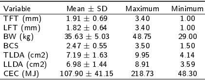
The study was carried out using data from twenty-two 3-year-old, non-pregnant, non-lactating Pelibuey ewes with body weight (BW) of 35.63 ± 5.03 kg (mean ± SD) and body condition score ± 0.55 (Table 1). BCS for each ewe was evaluated by two experienced technicians, using a 1-5 scale, with 0.5 increments, where BCS 1 represents a thin animal and 5 an obese animal as described by Russel et al. (1969).
Table 1. Mean (± SD) and minimum and maximum values for variables measured in adult Pelibuey ewes (n=22).

SD: standard deviation, BCS: body condition score, BW: body weight, TFT: thoracic fat thickness, LFT: lumbar fat thickness, TLDA: Thoracic L. dorsi area, LLDA: lumbar L. dorsi area, CEC: carcass energy content.
The ewes were selected from a commercial farm and were kept in roofed pens with a concrete floor and no walls. The diet consisted of 66 % forage and 34 % concéntrate, with an estimated metabolizable energy of 12 MJ KG-1 DM and 10 % CP (AFRC 1993). The dietary ingredients were cereal grains (corn or sorghum), soybean meal, hay tropical grasses, vitamins, and minerals.
USM were taken 24 h before slaughter. Fat thickness (FT) and Longissimus dorsi area (LDA) were determined using Pie Medical® 100 B-mode real-time ultrasound equipment, with a 6/8 MHz linear probe (Aguilar-Hernandez et al. 2016, Chay-Canul et al. 2016). Previously, the ewes were shaved between the 12th and 13th thoracic vertebrae (TFT and TLDA) and the 3rd and 4th lumbar vertebrae (LFT and LLDA) as described by Aguilar-Hernandez et al. (2016) and Chay-Canul et al. (2016). All measurements were taken on the left side of ewes and the area of the muscle (TLDA and LLDA) and fat thickness (TFT and LFT) in both regions were measured using the equipment’s digital callipers (Chay-Canul et al. 2016). USM were recorded on all animals using the same method described by Aguilar-Hernandez et al. (2016).
Slaughter of animals
The animals were humanely slaughtered following the Official Mexican standards (NOM-OS-ZOO, NOM-09-ZOO and NOM-033-ZOO) established for the slaughtering and processing of meat animals. Before slaughter, shrunk BW (SBW) was measured after feed and water were withdrawn for 24 h. The limbs, pelt, head and all internal organs were separated. The data recorded at slaughter were internal organs and hot carcass weights. Internal fat (TIF, internal adipose tissue) was dissected, weighed and grouped as either pelvic (around kidneys and pelvic región) or omental and mesenteric fat. Then the carcasses were split at the level of the dorsal midline in two equal halves, weighed, and chilled at 6 °C for 24 h. Subsequently, the left half-carcass was completely dissected into subcutaneous and intermuscular fat (carcass fat, CF), muscle, bone plus cartilage and each component was weighed separately. Dissected tissues of the left carcass were adjusted as whole carcass.
Chemical determinations
A sample of 500 g was taken from each animal from the muscle and adipose tissue of the carcass. This sample was ground separately with a screw grinder (Torrey, Co) through a 4 mm mesh; then the tissue samples were freeze-dried to determine water and dry matter content using freeze-dry equipment (Labconcco, Co., USA). The dry samples were then ground in a hammer mill for Chemical analyses for gross energy (adiabatic bomb calorimeter, Parr, USA). The CEC was determined as the sum of energy of muscle and adipose tissues in the carcass.
Data analysis
Correlation coefficients among variables were analysed by the PROC CORR procedure of SAS. Relationships between BW, BCS, USM and CEC were estimated by linear regression models using PROC REG. The STEPWISE option was used in the SELECTION statement for significant (p < 0.05) variables to be included in the statistical models. The accuracy of the models was evaluated by the determination coefficient (r2) and the mean square error (MSE).
Results and discussion
In an attempt to develop practical and inexpensive methods for predicting the Chemical composition (water, fat, protein and energy) of the body of farm animals, several methods have been evaluated, but some are very sophisticated, expensive and laborious. The USM method has shown great potential for this estimation. In hair sheep breeds, the use of USM is very limited. Although there are some studies that evaluated the use of USM to predict carcass traits, to our knowledge this is the first study that uses USM to assess the carcass energy content in Pelibuey ewes.
The means (± SD) and mínimum and máximum valúes for BW, BCS, USM and CEC for ewes used in this study are shown in Table 1. The correlation coefficient for BW and CEC was 0.89 (p < 0.001) and for BCS the r-value was 0.69. Nonetheless, the correlation between LDA (TLDA and LLDA) and CEC was low and not significant (Table 2). Conversely, for FT (TFT and LFT) the r-values ranged from 0.57 to 0.68 (p < 0.05).
Table 2. Correlation coefficients for body traits and carcass variables to predict the carcass energy content using ultrasound measurements in adult Pelibuey ewes in a tropical zone.

***P < 0.0001, **P < 0.001, *P < 0.05, BCS: body condition score, BW: body weight, TFT: thoracic fat thickness, LFT: lumbar fat thickness, TLDA: Thoracic L. dorsi area, LLDA: lumbar L. dorsi area, CEC: carcass energy content.
In the models selection, only the LFT was significant (p < 0.001). Simple regression equations using only the (LFT, Equation 1) had an r2 valué of 0.41 for CEC (RSD= 18.56). On the other hand, the inclusión of BW as an independent variable (Equation 2) increased the r2 valué to 0.79 (RSD= 18.88). The múltiple linear regression equation that included BW and LFT had the highest r2 valué (0.87; RSD= 15.34). The inclusión of LFT improved the prediction of carcass energy content by about 8 % (Table 3).
Table 3. Regression equations to predict the carcass energy content using ultrasound measurements in 22 adult Pelibuey ewes reared in a tropical zone.

R2: determination coefficient, MSE: mean square error, RSD: residual standard deviation, P: P-value.
In this sense, Silva et al. (2005), in working with lambs, reported that for predicting the carcass energy content, the subcutaneous fat depth over the 13th thoracic vertebra (TFT) explained 90 % of the variation, while the subcutaneous fat depth between the 3rd and the 4th lumbar vertebrae (LFT) explained 82 % of it. Also, Silva et al. (2016) observed similar results, reporting that the TFT explained around of 90 and 82 % of the variation in energy content of EBW in Churra da Terra Quente (CTQ) and lle de France (IF) ewes, respectively, while the LFT explained around 90 and 85 % of these variations in CTQ and IF ewes, respectively. In the current study the TFT was not significant in the models tested; moreover, the equations for predicting carcass energy content using LFT as the single predictor had an r2 that ranged from 0.41 to 0.47 (Equations 1, 2 and 3).
In the present study, the use of múltiple regression equations that included BW and LFT improved the accuracy of equations. The use of BW explained 60 to 80 % of the variation in carcass energy; moreover, the inclusión of LFT improves the prediction 5 to 15 %. BW, a trait easy to evaluate, is also a trait that in most studies of body composition tends to account for a large amount of the variation observed in different body components. This has been shown by Silva et al. (2005) and (Silva et al. 2016) in sheep, as in the current study.
In lambs, Silva et al. (2005) found that inclusión of BW and two of the three USM (TFT, LFT, and tissue depth over the 11th rib) explained 80 to 97 % (p < 0.01) of the carcass energy content and reported that the use of BW and some USM, particularly TFT, allow accurate prediction of carcass Chemical composition. The results of this study strongly suggest that body Chemical composition and the retained energy of adult Pelibuey ewes can be predicted by body weight and real-time ultra-sonography measurements. Also, Silva et al. (2016) found that múltiple regression models gave good estimates of Chemical body composition for CTQ and IF ewes from BW and USM.
The use of ultrasound measurements (LFT) in combination with BW in live adult Pelibuey ewes provided good estimates of the carcass energy content. It is important to know that ultrasound measurements can be used to estímate body composition of adult Pelibuey ewes because carcass information on these animals being evaluated for fattening purposes is not available.


 nueva página del texto (beta)


